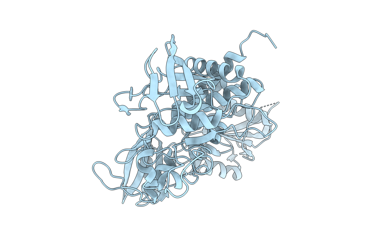
2B3O image
Deposition Date 2005-09-20
Release Date 2005-10-25
Last Version Date 2024-02-14
Entry Detail
PDB ID:
2B3O
Keywords:
Title:
Crystal structure of human tyrosine phosphatase SHP-1
Biological Source:
Source Organism:
Homo sapiens (Taxon ID: 9606)
Host Organism:
Method Details:
Experimental Method:
Resolution:
2.80 Å
R-Value Free:
0.29
R-Value Work:
0.23
R-Value Observed:
0.24
Space Group:
P 21 21 21
Macromolecular Entities
Polymer Type:polypeptide(L)
Molecule:Tyrosine-protein phosphatase, non-receptor type 6
Gene (Uniprot):PTPN6
Chain IDs:A
Chain Length:532
Number of Molecules:1
Biological Source:Homo sapiens
Primary Citation
Crystal structure of human protein-tyrosine phosphatase SHP-1.
J.Biol.Chem. 278 6516 6520 (2003)
PMID: 12482860 DOI: 10.1074/jbc.M210430200

Abstact

SHP-1 is a cytosolic protein-tyrosine phosphatase that behaves as a negative regulator in eukaryotic cellular signaling pathways. To understand its regulatory mechanism, we have determined the crystal structure of the C-terminal truncated human SHP-1 in the inactive conformation at 2.8-A resolution and refined the structure to a crystallographic R-factor of 24.0%. The three-dimensional structure shows that the ligand-free SHP-1 has an auto-inhibited conformation. Its N-SH2 domain blocks the catalytic domain and keeps the enzyme in the inactive conformation, which supports that the phosphatase activity of SHP-1 is primarily regulated by the N-SH2 domain. In addition, the C-SH2 domain of SHP-1 has a different orientation from and is more flexible than that of SHP-2, which enables us to propose an enzymatic activation mechanism in which the C-SH2 domains of SHPs could be involved in searching for phosphotyrosine activators.

Legend

Protein

Chemical

Disease

Primary Citation of related structures