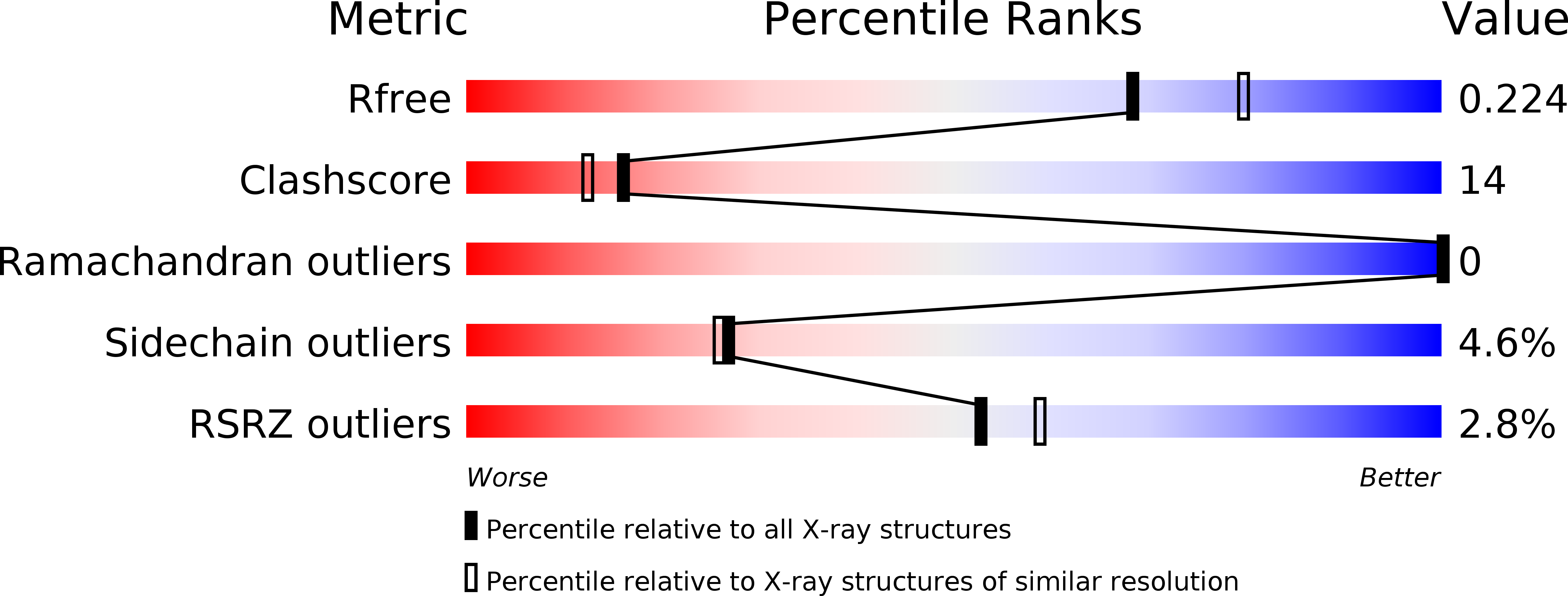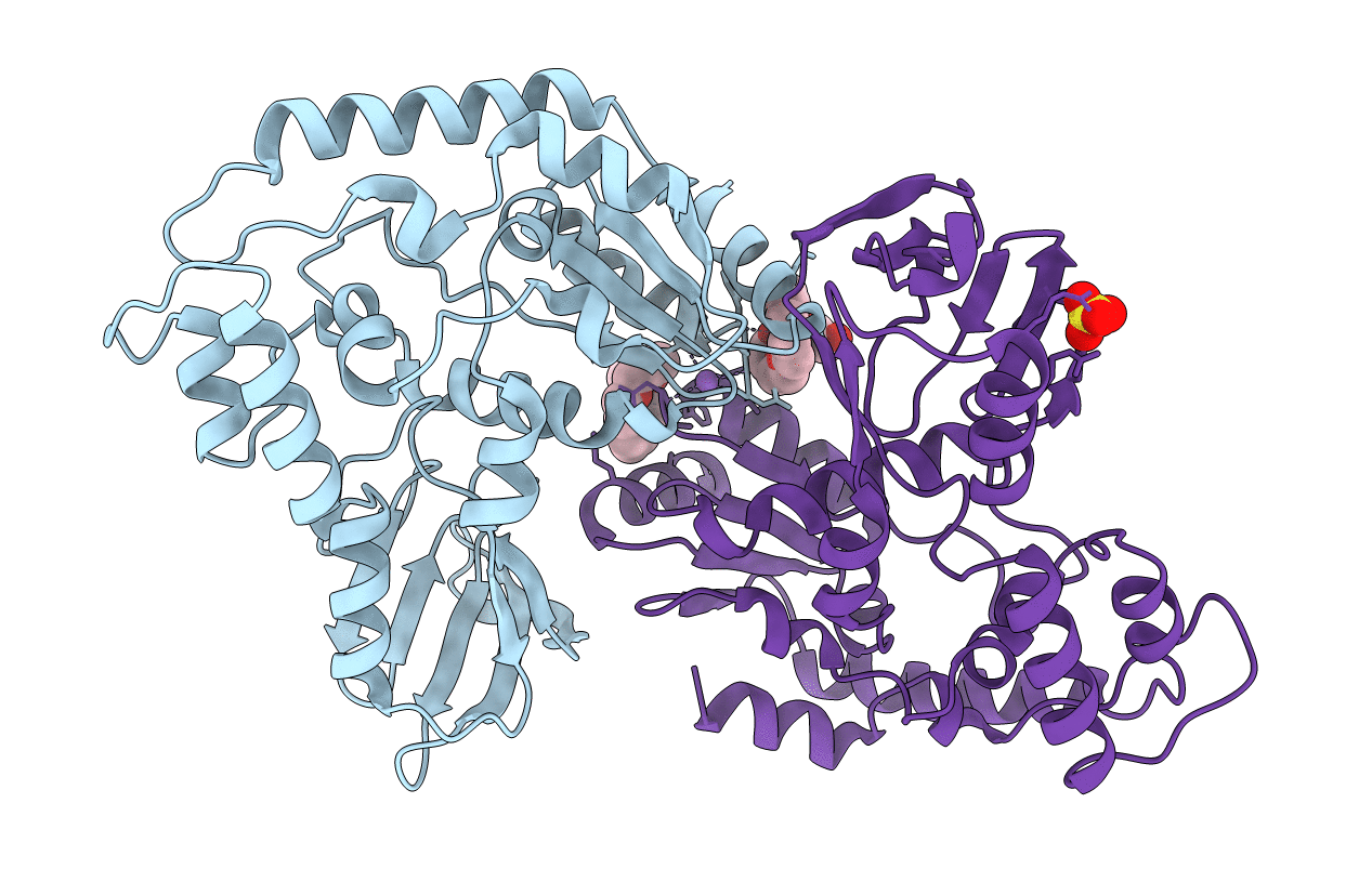
2B2N image
Deposition Date 2005-09-19
Release Date 2006-01-24
Last Version Date 2024-02-14
Entry Detail
PDB ID:
2B2N
Title:
Structure of transcription-repair coupling factor
Biological Source:
Source Organism:
Host Organism:
Method Details:
Experimental Method:
Resolution:
2.10 Å
R-Value Free:
0.23
R-Value Work:
0.19
Space Group:
P 65 2 2
Macromolecular Entities
Structures with similar UniProt ID
Protein Blast
Polymer Type:polypeptide(L)
Molecule:Transcription-repair coupling factor
Gene (Uniprot):mfd
Chain IDs:A, B
Chain Length:344
Number of Molecules:2
Biological Source:Escherichia coli
Primary Citation
Structural Basis for Transcription-coupled Repair: the N Terminus of Mfd Resembles UvrB with Degenerate ATPase Motifs
J.Mol.Biol. 355 675 683 (2006)
PMID: 16309703 DOI: 10.1016/j.jmb.2005.10.033

Abstact

The transcription repair coupling factor Mfd removes stalled RNA polymerase from DNA lesions and links transcription to UvrABC-dependent nucleotide excision repair in prokaryotes. We report the 2.1A crystal structure of the UvrA-binding N terminus (residues 1-333) of Escherichia coli Mfd (Mfd-N). Remarkably, Mfd-N reveals a fold that resembles the three N-terminal domains of the repair enzyme UvrB. Domain 1A of Mfd adopts a typical RecA fold, domain 1B matches the damage-binding domain of the UvrB, and domain 2 highly resembles the implicated UvrA-binding domain of UvrB. However, Mfd apparently lacks a functional ATP-binding site and does not contain the DNA damage-binding motifs of UvrB. Thus, our results suggest that Mfd might form a UvrA recruitment factor at stalled transcription complexes that architecturally but not catalytically resembles UvrB.

Legend

Protein

Chemical

Disease

Primary Citation of related structures