2B17 image
Entry Detail
PDB ID:
2B17
Keywords:
Title:
Specific binding of non-steroidal anti-inflammatory drugs (NSAIDs) to phospholipase A2: Crystal structure of the complex formed between phospholipase A2 and diclofenac at 2.7 A resolution:
Biological Source:
PDB Version:
Deposition Date:
2005-09-15
Release Date:
2005-10-04
Method Details:
Experimental Method:
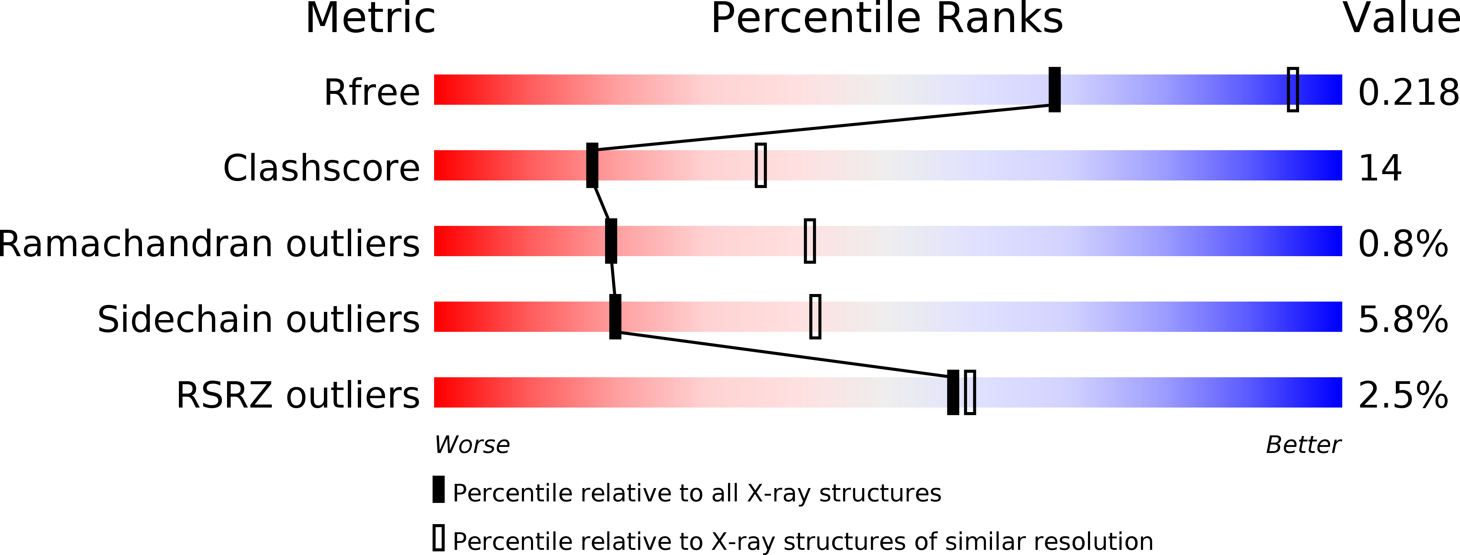
Resolution:
2.71 Å
R-Value Free:
0.21
R-Value Work:
0.19
R-Value Observed:
0.19
Space Group:
P 43
Macromolecular Entities
Polymer Type:polypeptide(L)
Description:Phospholipase A2 VRV-PL-VIIIa
Chain IDs:A
Chain Length:121
Number of Molecules:1
Biological Source:Daboia russellii pulchella
Primary Citation
Specific binding of non-steroidal anti-inflammatory drugs (NSAIDs) to phospholipase A2: structure of the complex formed between phospholipase A2 and diclofenac at 2.7 A resolution.
Acta Crystallogr.,Sect.D 62 410 416 (2006)
PMID: 16552142 DOI: 10.1107/S0907444906003660

Abstact

Type IIA secretory phospholipase A2 (PLA2) enzymes catalyze the hydrolysis of the sn-2 ester bond of glycerophospholipids to release fatty acids and lysophospholipids. In order to elucidate the role of PLA2 in inflammatory disorders and to determine the mode of binding of non-steroidal anti-inflammatory drugs (NSAIDs) to PLA2, the detailed three-dimensional structure of a complex formed between a group IIA PLA2 from Daboia russelli pulchella and 2-[(2,6-dichlorophenyl)amino]benzeneacetic acid (diclofenac) has been determined. The preformed complex was crystallized by equilibrating the protein solution against a mixture of 0.20 M ammonium sulfate and 30% PEG 4000. The crystals belong to space group P4(3), with unit-cell parameters a = b = 53.0, c = 48.4 A. The structure was solved by the molecular-replacement method and refined to R(cryst) and R(free) factors of 0.192 and 0.211, respectively, using reflections to 2.7 A resolution. The structure showed that diclofenac occupies a very favourable position in the centre of the substrate-binding hydrophobic channel that allows a number of intermolecular interactions. The binding mode of diclofenac involved crucial interactions with important residues for substrate recognition such as Asp49, His48 and Gly30. In addition, it included three new interactions involving its Cl atoms with Phe5, Ala18 and Tyr22. It also showed an extensive network of hydrophobic interactions involving almost all of the residues of the substrate-binding hydrophobic channel. The binding affinity of diclofenac was determined using surface plasmon resonance, which gave an equilibrium constant of 4.8 +/- 0.2 x 10(-8) M.

Legend

Protein

Chemical

Disease

Primary Citation of related structures