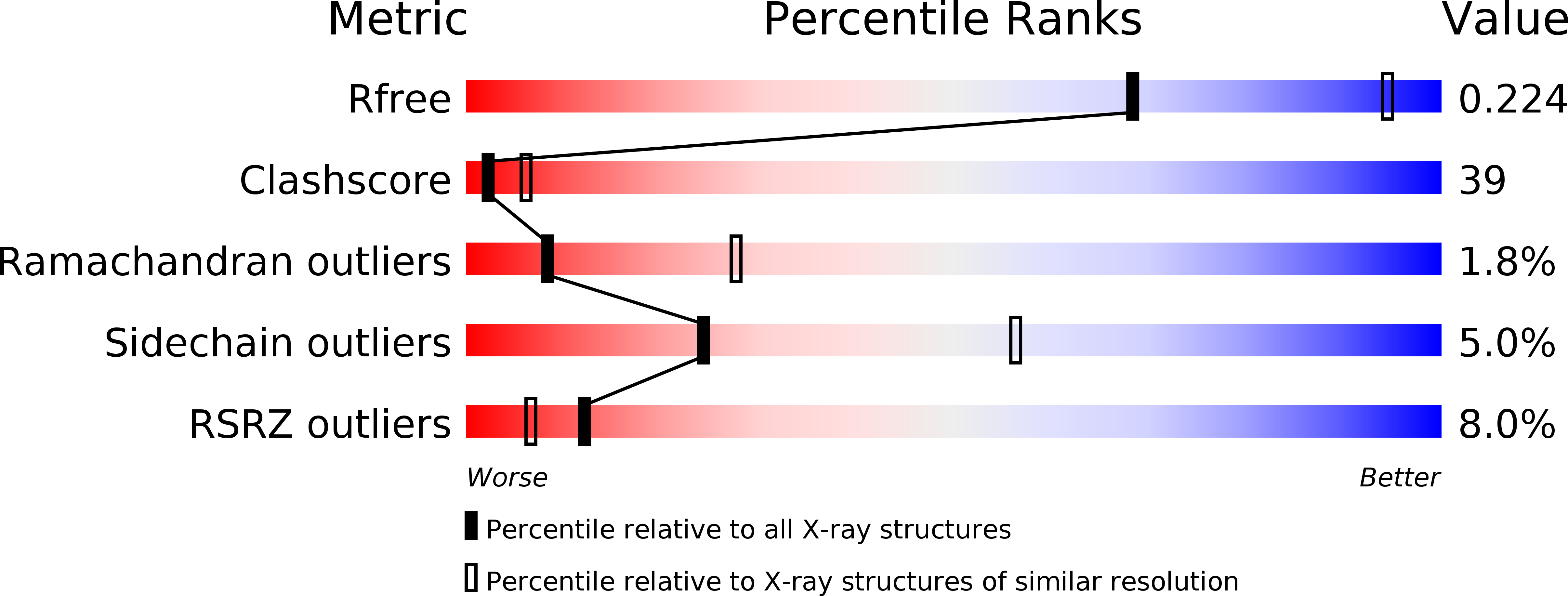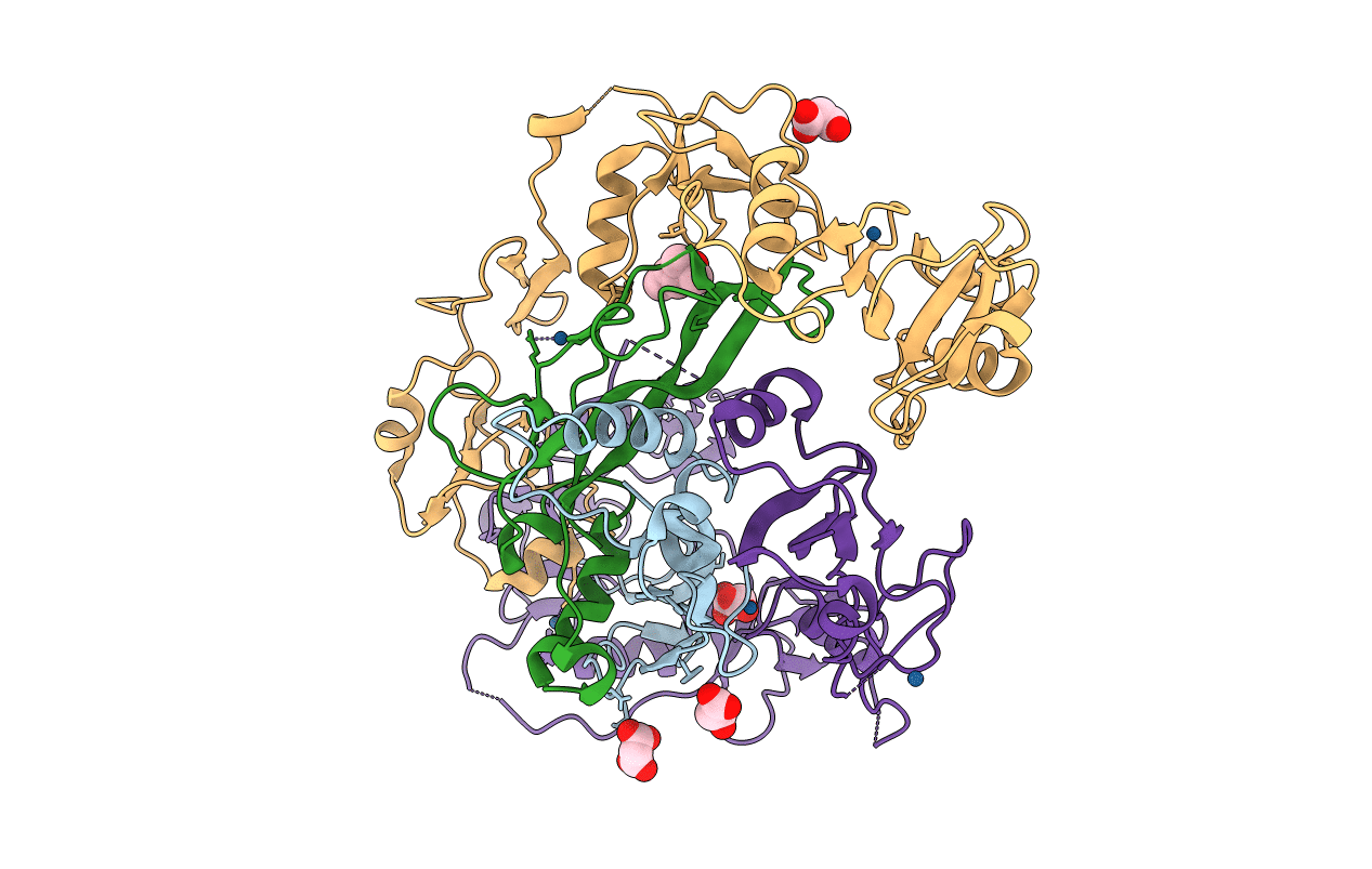
2B0U image
Entry Detail
PDB ID:
2B0U
Title:
The Structure of the Follistatin:Activin Complex
Biological Source:
Source Organism:
Host Organism:
PDB Version:
Deposition Date:
2005-09-14
Release Date:
2005-10-11
Method Details:
Experimental Method:
Resolution:
2.80 Å
R-Value Free:
0.29
R-Value Work:
0.26
R-Value Observed:
0.30
Space Group:
P 21 21 2
Macromolecular Entities
Polymer Type:polypeptide(L)
Description:Inhibin beta A chain
Chain IDs:A, B
Chain Length:116
Number of Molecules:2
Biological Source:Homo sapiens
Polymer Type:polypeptide(L)
Description:Follistatin
Chain IDs:C, D
Chain Length:288
Number of Molecules:2
Biological Source:Homo sapiens
Primary Citation
The Structure of the Follistatin:Activin Complex Reveals Antagonism of Both Type I and Type II Receptor Binding.
Dev.Cell 9 535 543 (2005)
PMID: 16198295 DOI: 10.1016/j.devcel.2005.09.008

Abstact

TGF-beta ligands stimulate diverse cellular differentiation and growth responses by signaling through type I and II receptors. Ligand antagonists, such as follistatin, block signaling and are essential regulators of physiological responses. Here we report the structure of activin A, a TGF-beta ligand, bound to the high-affinity antagonist follistatin. Two follistatin molecules encircle activin, neutralizing the ligand by burying one-third of its residues and its receptor binding sites. Previous studies have suggested that type I receptor binding would not be blocked by follistatin, but the crystal structure reveals that the follistatin N-terminal domain has an unexpected fold that mimics a universal type I receptor motif and occupies this receptor binding site. The formation of follistatin:BMP:type I receptor complexes can be explained by the stoichiometric and geometric arrangement of the activin:follistatin complex. The mode of ligand binding by follistatin has important implications for its ability to neutralize homo- and heterodimeric ligands of this growth factor family.

Legend

Protein

Chemical

Disease

Primary Citation of related structures