Deposition Date
1991-01-11
Release Date
1993-07-15
Last Version Date
2024-11-20
Entry Detail
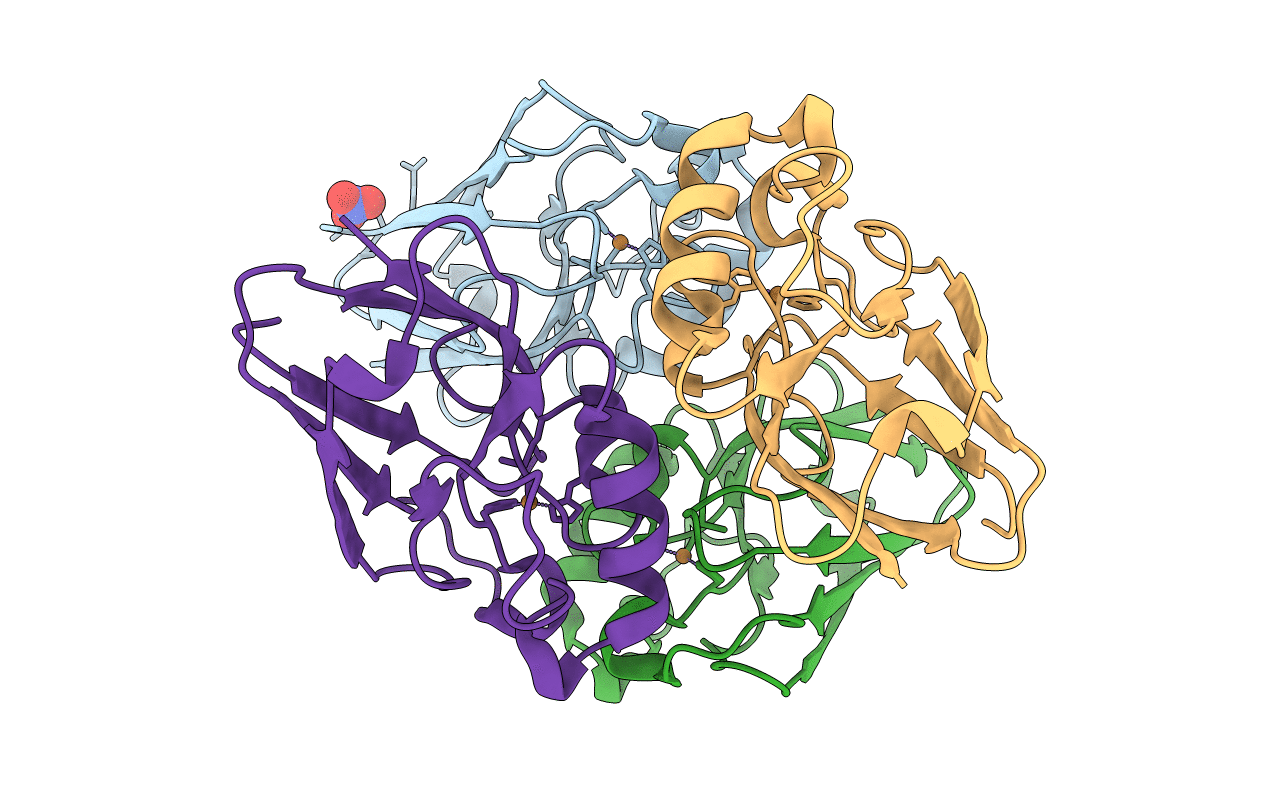
PDB ID:
2AZU
Keywords:
Title:
X-RAY CRYSTAL STRUCTURE OF THE TWO SITE-SPECIFIC MUTANTS HIS35*GLN AND HIS35*LEU OF AZURIN FROM PSEUDOMONAS AERUGINOSA
Biological Source:
Source Organism(s):
Pseudomonas aeruginosa (Taxon ID: 287)
Method Details:
Experimental Method:
Resolution:
1.90 Å
R-Value Work:
0.17
Space Group:
P 21 21 21