2AYL image
Entry Detail
PDB ID:
2AYL
Keywords:
Title:
2.0 Angstrom Crystal Structure of Manganese Protoporphyrin IX-reconstituted Ovine Prostaglandin H2 Synthase-1 Complexed With Flurbiprofen
Biological Source:
Source Organism:
PDB Version:
Deposition Date:
2005-09-07
Release Date:
2006-01-24
Method Details:
Experimental Method:
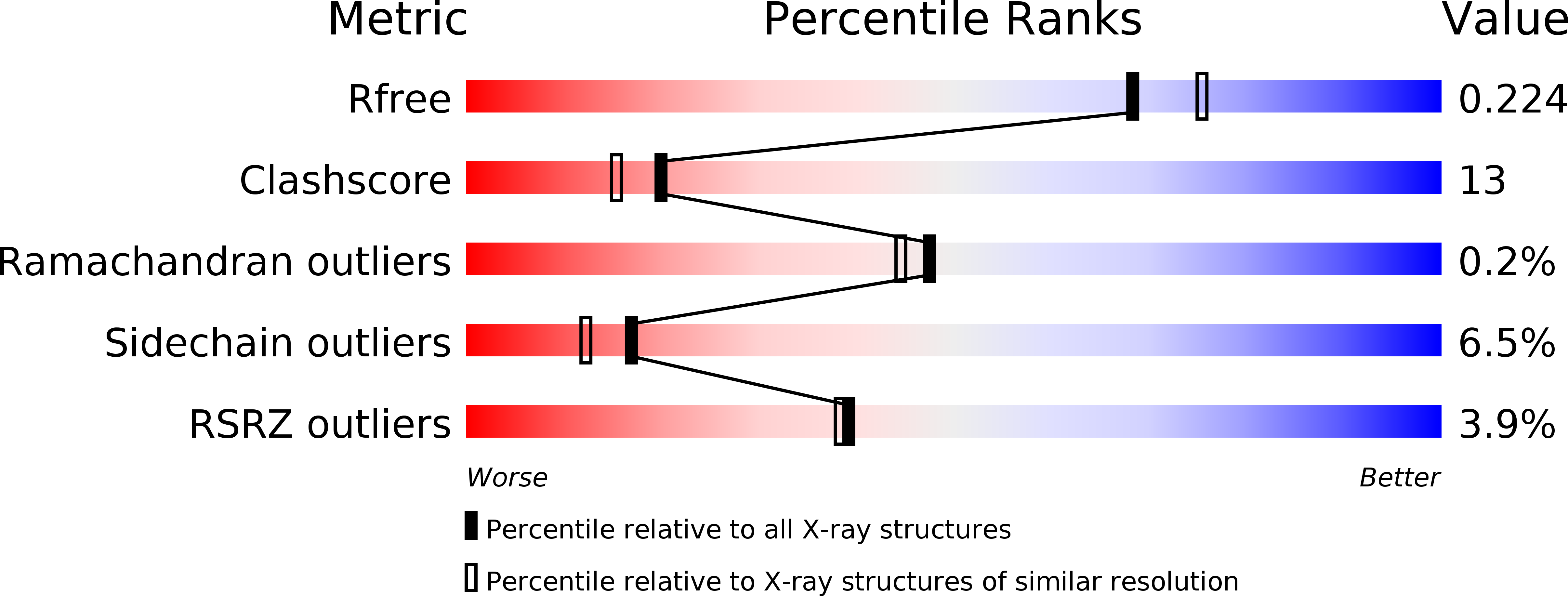
Resolution:
2.00 Å
R-Value Free:
0.23
R-Value Work:
0.21
Space Group:
I 2 2 2
Macromolecular Entities
Polymer Type:polypeptide(L)
Description:Prostaglandin G/H synthase 1
Chain IDs:A, B
Chain Length:553
Number of Molecules:2
Biological Source:Ovis aries
Modified Residue
Compound ID Chain ID Parent Comp ID Details 2D Image
ASN A ASN GLYCOSYLATION SITE
Primary Citation
2.0 angstroms structure of prostaglandin H2 synthase-1 reconstituted with a manganese porphyrin cofactor.
Acta Crystallogr.,Sect.D 62 151 156 (2006)
PMID: 16421446 DOI: 10.1107/S0907444905036309

Abstact

Prostaglandin H2 synthase (EC 1.14.99.1) is a clinically important drug target that catalyzes two key steps in the biosynthesis of the eicosanoid hormones. The enzyme contains spatially distinct cyclooxygenase and peroxidase active sites, both of which require a heme cofactor. Substitution of ferric heme by Mn(III) protoporphyrin IX greatly diminishes the peroxidase activity, but has little effect on the cyclooxygenase activity. Here, the 2.0 angstroms resolution crystal structure of the Mn(III) form of ovine prostaglandin H2 synthase-1 is described (R = 21.8%, R(free) = 23.7%). Substitution of Mn(III) for Fe(III) causes no structural perturbations in the protein. However, the out-of-plane displacement of the manganese ion with respect to the porphyrin is greater than that of the iron by approximately 0.2 angstroms. This perturbation may help to explain the altered catalytic properties of the manganese enzyme.

Legend

Protein

Chemical

Disease

Primary Citation of related structures