Deposition Date
2005-09-06
Release Date
2006-08-15
Last Version Date
2024-02-14
Entry Detail
PDB ID:
2AY0
Keywords:
Title:
Structure of the Lys9Met mutant of the E. coli Proline Utilization A (PutA) DNA-binding domain.
Biological Source:
Source Organism(s):
Escherichia coli (Taxon ID: 562)
Expression System(s):
Method Details:
Experimental Method:
Resolution:
2.10 Å
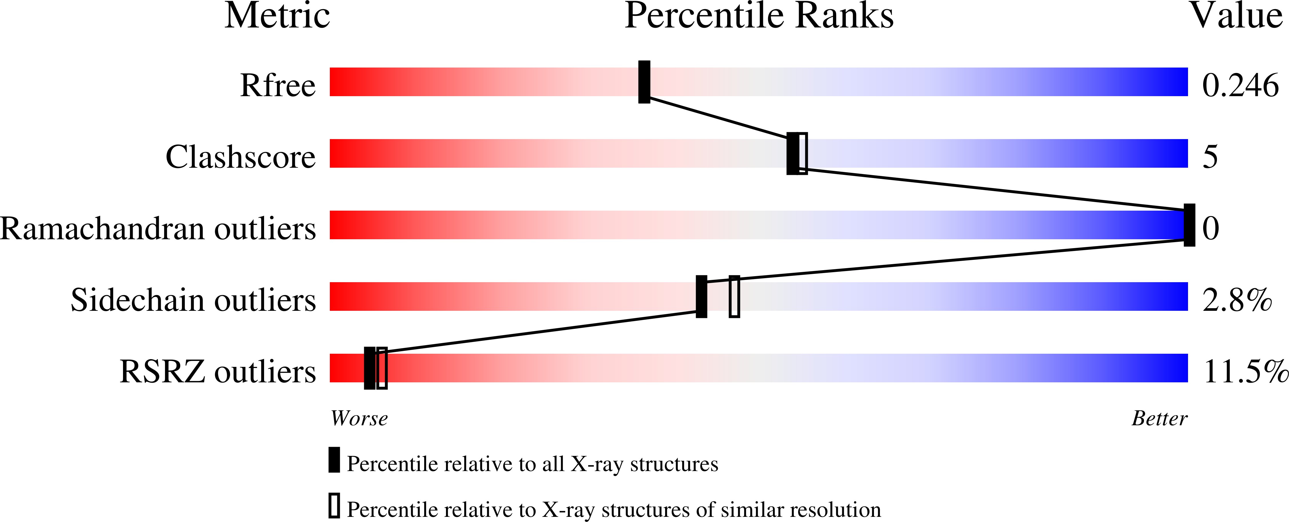
R-Value Free:
0.24
R-Value Work:
0.20
R-Value Observed:
0.20
Space Group:
C 1 2 1