2AXW image
Deposition Date 2005-09-06
Release Date 2005-11-01
Last Version Date 2024-11-20
Entry Detail
PDB ID:
2AXW
Keywords:
Title:
Structure of DraD invasin from uropathogenic Escherichia coli
Biological Source:
Source Organism:
Escherichia coli (Taxon ID: 562)
Host Organism:
Method Details:
Experimental Method:
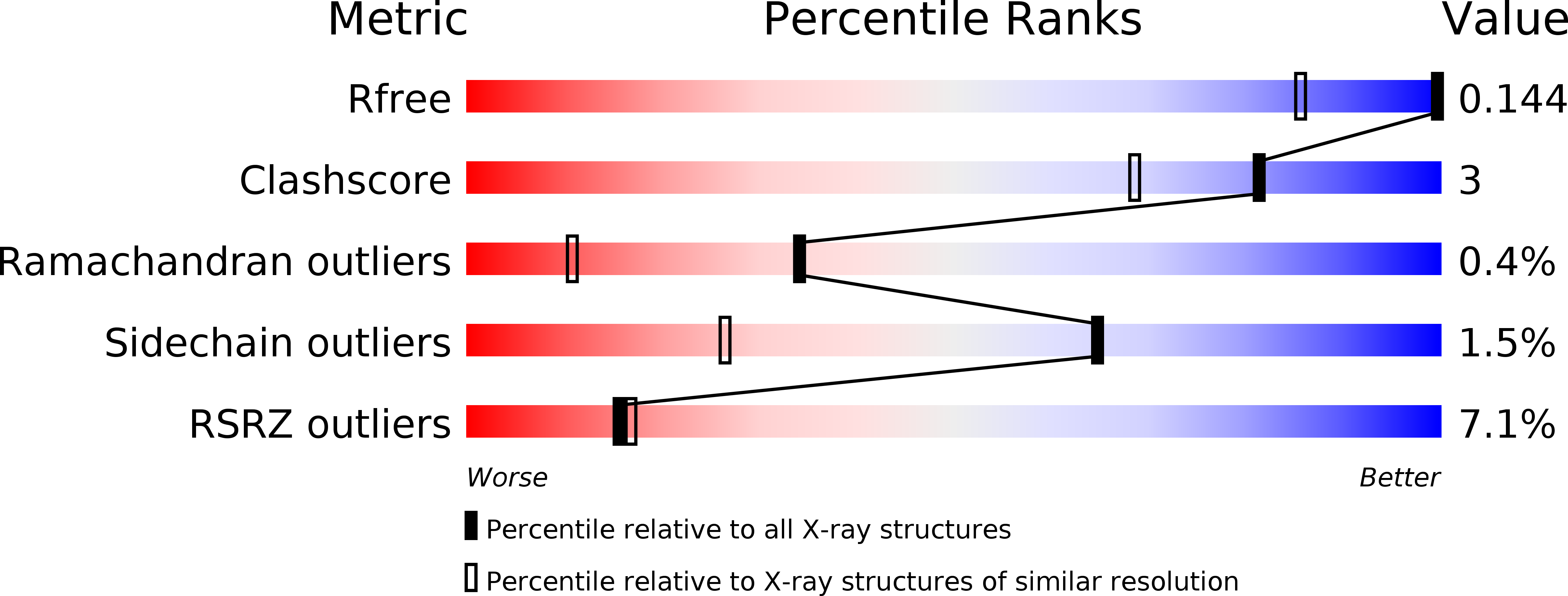
Resolution:
1.05 Å
R-Value Free:
0.16
R-Value Work:
0.15
R-Value Observed:
0.15
Space Group:
P 21 21 21
Macromolecular Entities
Structures with similar UniProt ID
Protein Blast
Polymer Type:polypeptide(L)
Molecule:DraD invasin
Chain IDs:A, B
Chain Length:134
Number of Molecules:2
Biological Source:Escherichia coli
Primary Citation
Structure of DraD invasin from uropathogenic Escherichia coli: a dimer with swapped beta-tails.
Acta Crystallogr.,Sect.D 62 157 164 (2006)
PMID: 16421447 DOI: 10.1107/S0907444905036747

Abstact

The dra gene cluster of uropathogenic strains of Escherichia coli produces proteins involved in bacterial attachment to and invasion of the eukaryotic host tissues. The crystal structure of a construct of E. coli DraD possessing an additional C-terminal extension of 13 amino acids, including a His6 tag, has been solved at a resolution of 1.05 angstroms. The protein forms symmetric dimers through the exchange of the C-terminal beta-strands, which participate in the immunoglobulin-like beta-sandwich fold of each subunit. This structure confirms that DraD is able to act as an acceptor in the donor-strand complementation mechanism of fiber formation but, in contrast to DraE adhesin, its native sequence does not have a donor strand; therefore, DraD can only be located at the tip of the fiber.

Legend

Protein

Chemical

Disease

Primary Citation of related structures