2AXR image
Deposition Date 2005-09-05
Release Date 2005-09-13
Last Version Date 2024-11-20
Entry Detail
PDB ID:
2AXR
Keywords:
Title:
Crystal structure of glucooligosaccharide oxidase from Acremonium strictum: a novel flavinylation of 6-S-cysteinyl, 8alpha-N1-histidyl FAD
Biological Source:
Source Organism:
Host Organism:
Method Details:
Experimental Method:
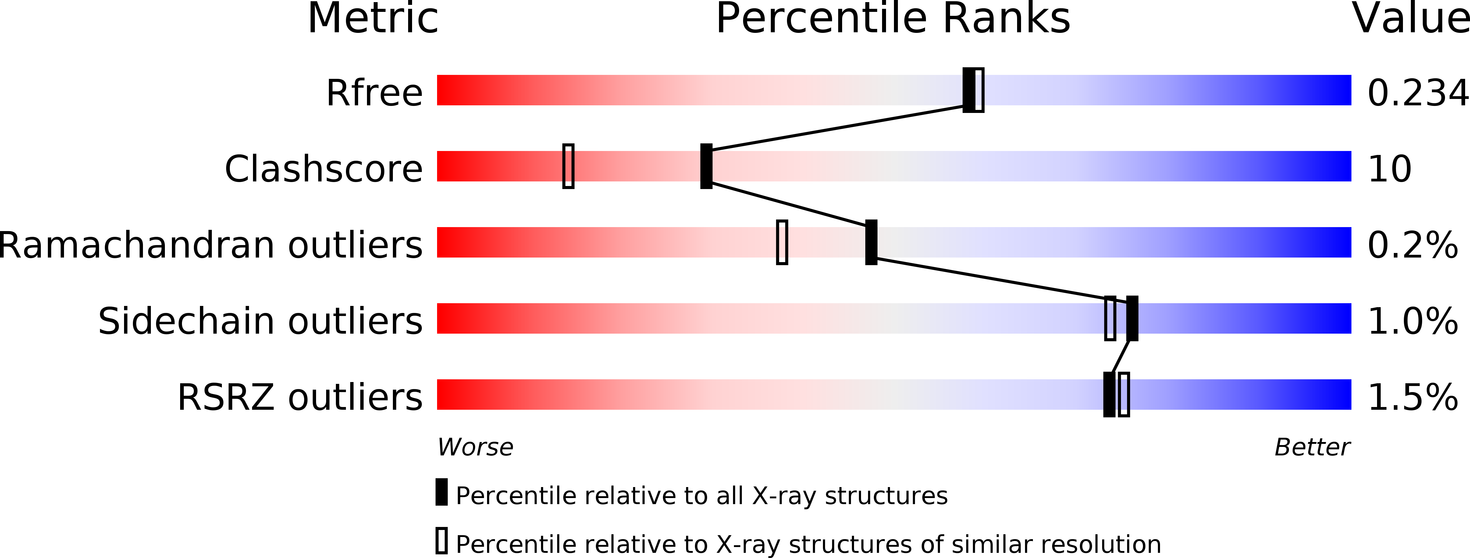
Resolution:
1.98 Å
R-Value Free:
0.24
R-Value Work:
0.19
R-Value Observed:
0.20
Space Group:
P 21 21 21
Macromolecular Entities
Polymer Type:polypeptide(L)
Molecule:glucooligosaccharide oxidase
Gene (Uniprot):gluO
Chain IDs:A
Chain Length:503
Number of Molecules:1
Biological Source:Acremonium strictum
Modified Residue
Compound ID Chain ID Parent Comp ID Details 2D Image
ASN A ASN GLYCOSYLATION SITE
Primary Citation
Crystal Structure of Glucooligosaccharide Oxidase from Acremonium strictum: A NOVEL FLAVINYLATION OF 6-S-CYSTEINYL, 8{alpha}-N1-HISTIDYL FAD
J.Biol.Chem. 280 38831 38838 (2005)
PMID: 16154992 DOI: 10.1074/jbc.M506078200

Abstact

Glucooligosaccharide oxidase from Acremonium strictum has been screened for potential applications in oligosaccharide acid production and alternative carbohydrate detection, because it catalyzes the oxidation of glucose, maltose, lactose, cellobiose and cello- and maltooligosaccharides. We report the crystal structures of the enzyme and of its complex with an inhibitor, 5-amino-5-deoxy- cellobiono-1,5-lactam at 1.55- and 1.98-A resolution, respectively. Unexpectedly, the protein structure demonstrates the first known double attachment flavinylation, 6-S-cysteinyl, 8alpha-N1-histidyl FAD. The FAD cofactor is cross-linked to the enzyme via the C(6) atom and the 8alpha-methyl group of the isoalloxazine ring with Cys(130) and His(70), respectively. This sugar oxidase possesses an open carbohydrate-binding groove, allowing the accommodation of higher oligosaccharides. The complex structure suggests that this enzyme may prefer a beta-d-glucosyl residue at the reducing end with the conserved Tyr(429) acting as a general base to abstract the OH(1) proton in concert with the H(1) hydride transfer to the flavin N(5). Finally, a detailed comparison illustrates the structural conservation as well as the divergence between this protein and its related flavoenzymes.

Legend

Protein

Chemical

Disease

Primary Citation of related structures