2AXQ image
Deposition Date 2005-09-05
Release Date 2006-08-15
Last Version Date 2023-08-23
Entry Detail
PDB ID:
2AXQ
Keywords:
Title:
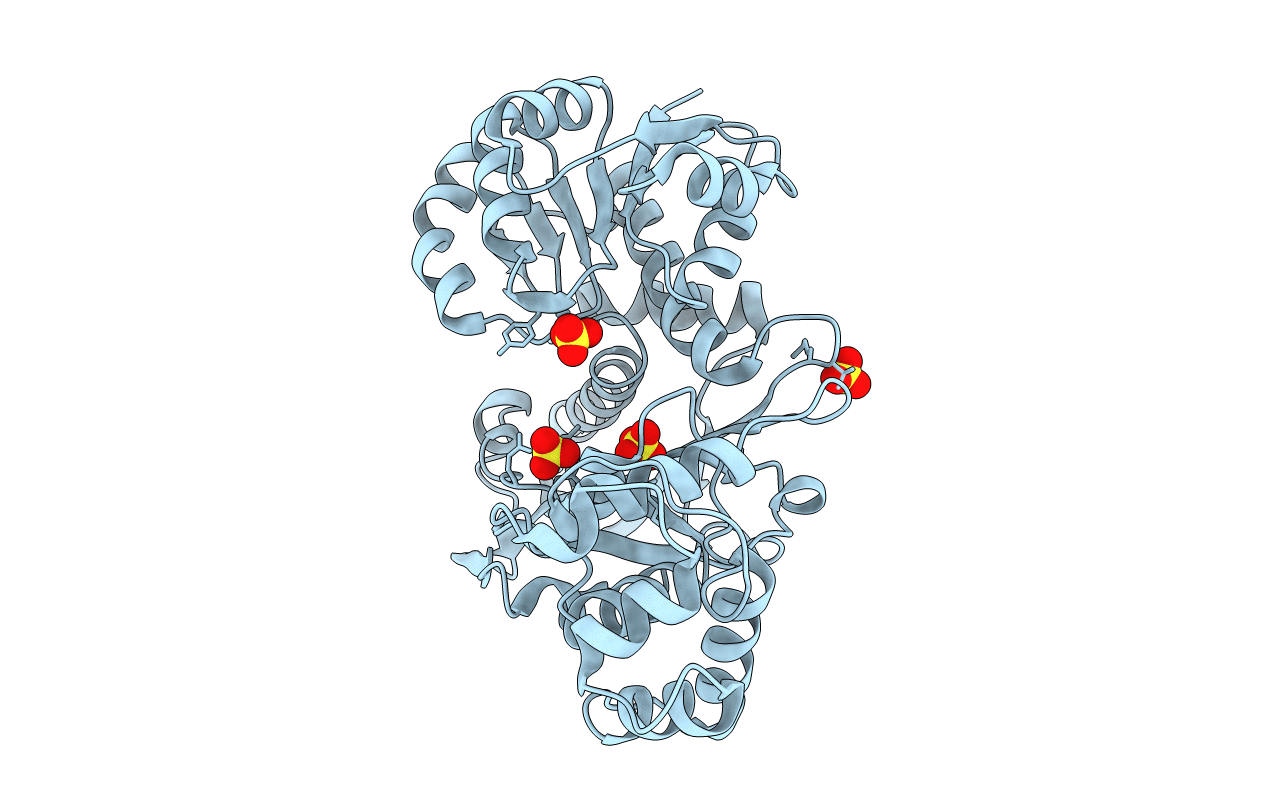
Apo histidine-tagged saccharopine dehydrogenase (L-Glu forming) from Saccharomyces cerevisiae
Biological Source:
Source Organism:
Host Organism:
Method Details:
Experimental Method:
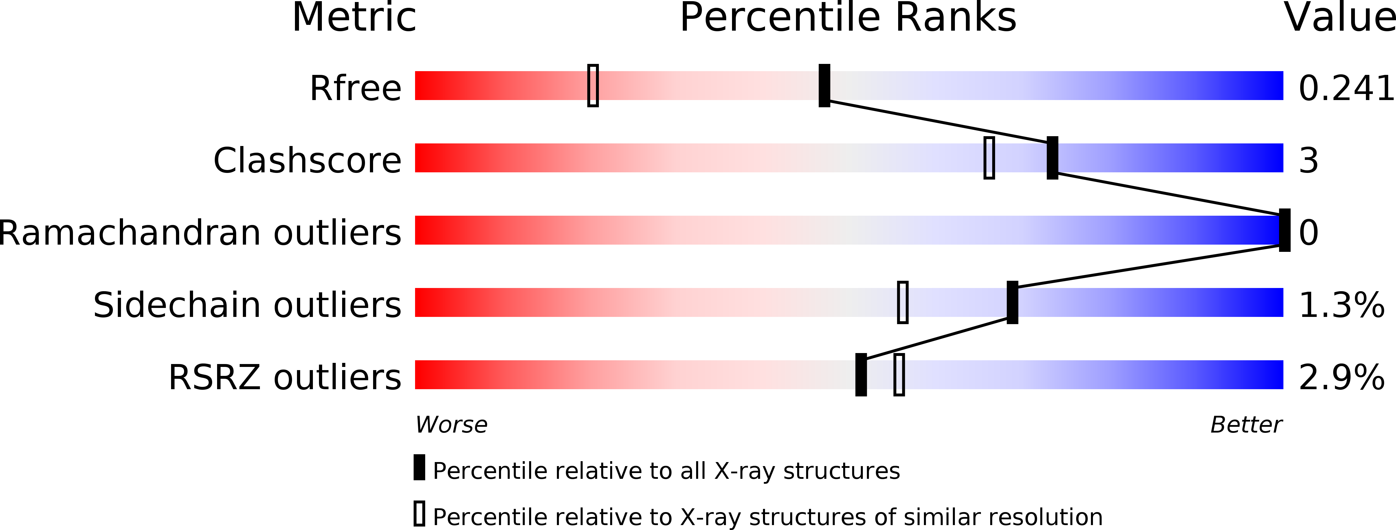
Resolution:
1.70 Å
R-Value Free:
0.24
R-Value Work:
0.19
R-Value Observed:
0.2
Space Group:
P 31 2 1
Macromolecular Entities
Polymer Type:polypeptide(L)
Molecule:Saccharopine dehydrogenase
Gene (Uniprot):LYS9
Chain IDs:A
Chain Length:467
Number of Molecules:1
Biological Source:Saccharomyces cerevisiae
Ligand Molecules
Primary Citation
Crystal structure of the his-tagged saccharopine reductase from Saccharomyces cerevisiae at 1.7-A resolution.
Cell Biochem.Biophys. 46 17 26 (2006)
PMID: 16943620 DOI: 10.1385/CBB:46:1:17

Abstact

The three-dimensional structure of the saccharopine reductase enzyme from the budding yeast Saccharomyces cerevisiae was determined to 1.7-A resolution in the apo form by using molecular replacement. The enzyme monomer consists of three domains: domain I is a variant of the Rossmann fold, domain II folds into a alpha/beta structure containing a mixed seven-stranded beta-sheet as the central core, and domain III has an all-helical fold. Comparative fold alignment with the enzyme from Magnaporthe grisea suggests that domain I binds to NADPH, and domain II binds to saccharopine and is involved in dimer formation. Domain III is involved in closing the active site of the enzyme once substrates are bound. Structural comparison of the saccharopine reductase enzymes from S. cerevisiae and M. grisea indicates that domain II has the highest number of conserved residues, suggesting that it plays an important role in substrate binding and in spatially orienting domains I and III.

Legend

Protein

Chemical

Disease

Primary Citation of related structures