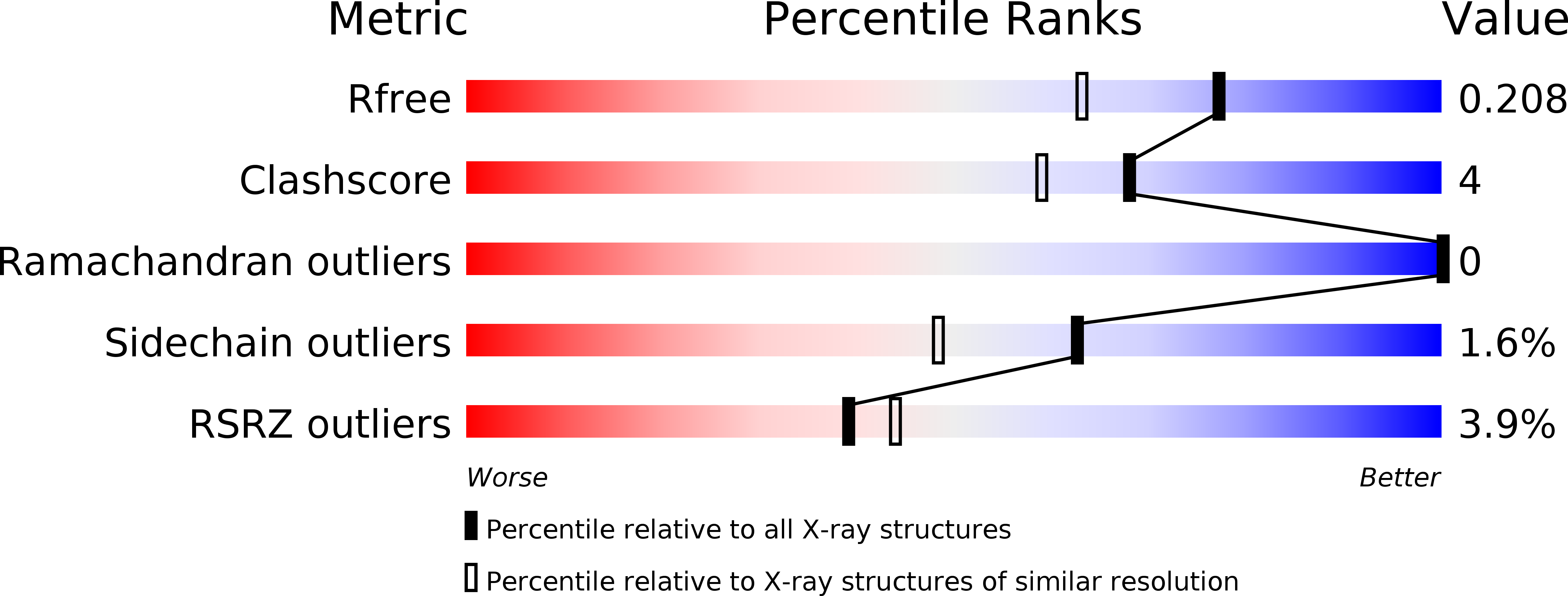
2AXC image
Deposition Date 2005-09-04
Release Date 2006-03-14
Last Version Date 2024-10-23
Entry Detail
PDB ID:
2AXC
Keywords:
Title:
Crystal structure of ColE7 translocation domain
Biological Source:
Source Organism:
Host Organism:
Method Details:
Experimental Method:
Resolution:
1.70 Å
R-Value Free:
0.20
R-Value Work:
0.18
R-Value Observed:
0.18
Space Group:
P 31 2 1
Macromolecular Entities
Polymer Type:polypeptide(L)
Molecule:Colicin E7
Gene (Uniprot):colE7
Chain IDs:A
Chain Length:264
Number of Molecules:1
Biological Source:Escherichia coli str. K12 substr.
Modified Residue
Compound ID Chain ID Parent Comp ID Details 2D Image
MSE A MET SELENOMETHIONINE
Primary Citation
High-resolution crystal structure of a truncated ColE7 translocation domain: implications for colicin transport across membranes
J.Mol.Biol. 356 22 31 (2006)
PMID: 16360169 DOI: 10.1016/j.jmb.2005.11.056

Abstact

ColE7 is a nuclease-type colicin released from Escherichia coli to kill sensitive bacterial cells by degrading the nucleic acid molecules in their cytoplasm. ColE7 is classified as one of the group A colicins, since the N-terminal translocation domain (T-domain) of the nuclease-type colicins interact with specific membrane-bound or periplasmic Tol proteins during protein import. Here, we show that if the N-terminal tail of ColE7 is deleted, ColE7 (residues 63-576) loses its bactericidal activity against E.coli. Moreover, TolB protein interacts directly with the T-domain of ColE7 (residues 1-316), but not with the N-terminal deleted T-domain (residues 60-316), as detected by co-immunoprecipitation experiments, confirming that the N-terminal tail is required for ColE7 interactions with TolB. The crystal structure of the N-terminal tail deleted ColE7 T-domain was determined by the multi-wavelength anomalous dispersion method at a resolution of 1.7 angstroms. The structure of the ColE7 T-domain superimposes well with the T-domain of ColE3 and TR-domain of ColB, a group A Tol-dependent colicin and a group B TonB-dependent colicin, respectively. The structural resemblance of group A and B colicins implies that the two groups of colicins may share a mechanistic connection during cellular import.

Legend

Protein

Chemical

Disease

Primary Citation of related structures