2AV5 image
Deposition Date 2005-08-29
Release Date 2006-01-24
Last Version Date 2024-02-14
Entry Detail
PDB ID:
2AV5
Keywords:
Title:
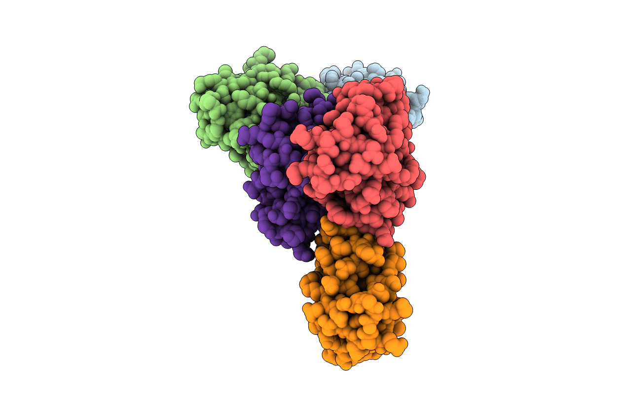
Crystal structure of Pyrococcus furiosus Pop5, an archaeal Ribonuclease P protein
Biological Source:
Source Organism:
Method Details:
Experimental Method:
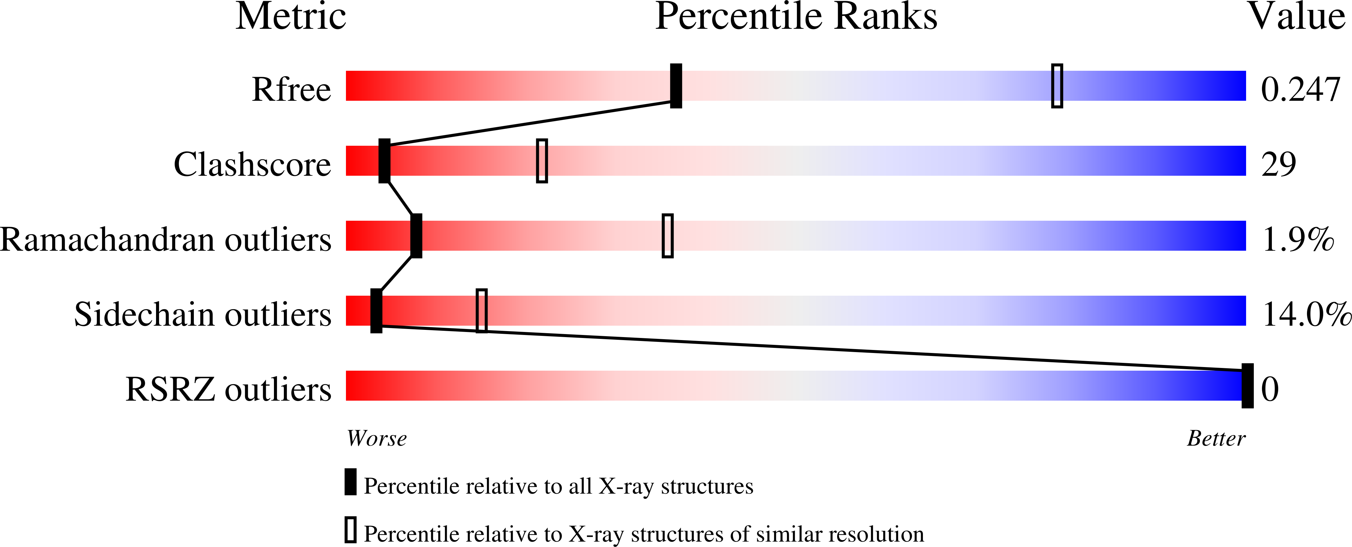
Resolution:
3.15 Å
R-Value Free:
0.26
R-Value Work:
0.23
R-Value Observed:
0.23
Space Group:
P 41 21 2
Macromolecular Entities
Polymer Type:polypeptide(L)
Molecule:Ribonuclease P protein component 2
Gene (Uniprot):rnp2
Mutations:C72S
Chain IDs:A, B, C, D, E
Chain Length:120
Number of Molecules:5
Biological Source:Pyrococcus furiosus
Ligand Molecules
Primary Citation
Structure of Pfu Pop5, an archaeal RNase P protein.
Proc.Natl.Acad.Sci.Usa 103 873 878 (2006)
PMID: 16418270 DOI: 10.1073/pnas.0508004103

Abstact

We have used NMR spectroscopy and x-ray crystallography to determine the three-dimensional structure of PF1378 (Pfu Pop5), one of four protein subunits of archaeal RNase P that shares a homolog in the eukaryotic enzyme. RNase P is an essential and ubiquitous ribonucleoprotein enzyme required for maturation of tRNA. In bacteria, the enzyme's RNA subunit is responsible for cleaving the single-stranded 5' leader sequence of precursor tRNA molecules (pre-tRNA), whereas the protein subunit assists in substrate binding. Although in bacteria the RNase P holoenzyme consists of one large catalytic RNA and one small protein subunit, in archaea and eukarya the enzyme contains several (> or =4) protein subunits, each of which lacks sequence similarity to the bacterial protein. The functional role of the proteins is poorly understood, as is the increased complexity in comparison to the bacterial enzyme. Pfu Pop5 has been directly implicated in catalysis by the observation that it pairs with PF1914 (Pfu Rpp30) to functionally reconstitute the catalytic domain of the RNA subunit. The protein adopts an alpha-beta sandwich fold highly homologous to the single-stranded RNA binding RRM domain. Furthermore, the three-dimensional arrangement of Pfu Pop5's structural elements is remarkably similar to that of the bacterial protein subunit. NMR spectra have been used to map the interaction of Pop5 with Pfu Rpp30. The data presented permit tantalizing hypotheses regarding the role of this protein subunit shared by archaeal and eukaryotic RNase P.

Legend

Protein

Chemical

Disease

Primary Citation of related structures