
Deposition Date
2005-08-29
Release Date
2005-10-18
Last Version Date
2024-10-09
Entry Detail
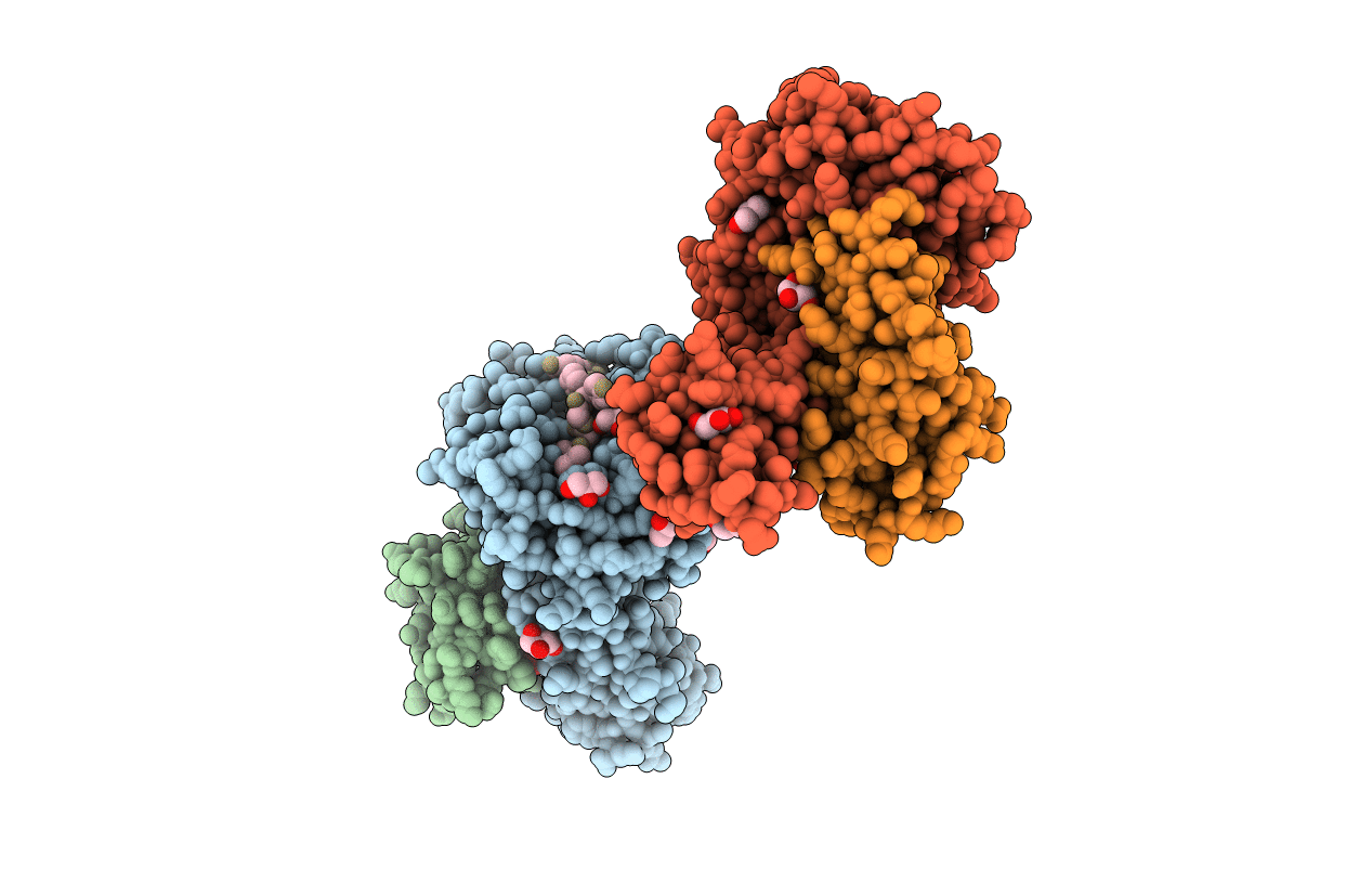
PDB ID:
2AV1
Keywords:
Title:
Crystal structure of HTLV-1 TAX peptide Bound to Human Class I MHC HLA-A2 with the E63Q and K66A mutations in the heavy chain.
Biological Source:
Source Organism(s):
Homo sapiens (Taxon ID: 9606)
Human T-cell leukemia virus 1 (isolate Caribbea HS-35 subtype A) (Taxon ID: 11927)
Human T-cell leukemia virus 1 (isolate Caribbea HS-35 subtype A) (Taxon ID: 11927)
Expression System(s):
Method Details:
Experimental Method:
Resolution:
1.95 Å
R-Value Free:
0.23
R-Value Work:
0.17
R-Value Observed:
0.17
Space Group:
P 1
