2AUC image
Deposition Date 2005-08-27
Release Date 2006-01-17
Last Version Date 2025-03-26
Entry Detail
PDB ID:
2AUC
Title:
Structure of the Plasmodium MTIP-MyoA complex, a key component of the parasite invasion motor
Biological Source:
Source Organism:
Host Organism:
Method Details:
Experimental Method:
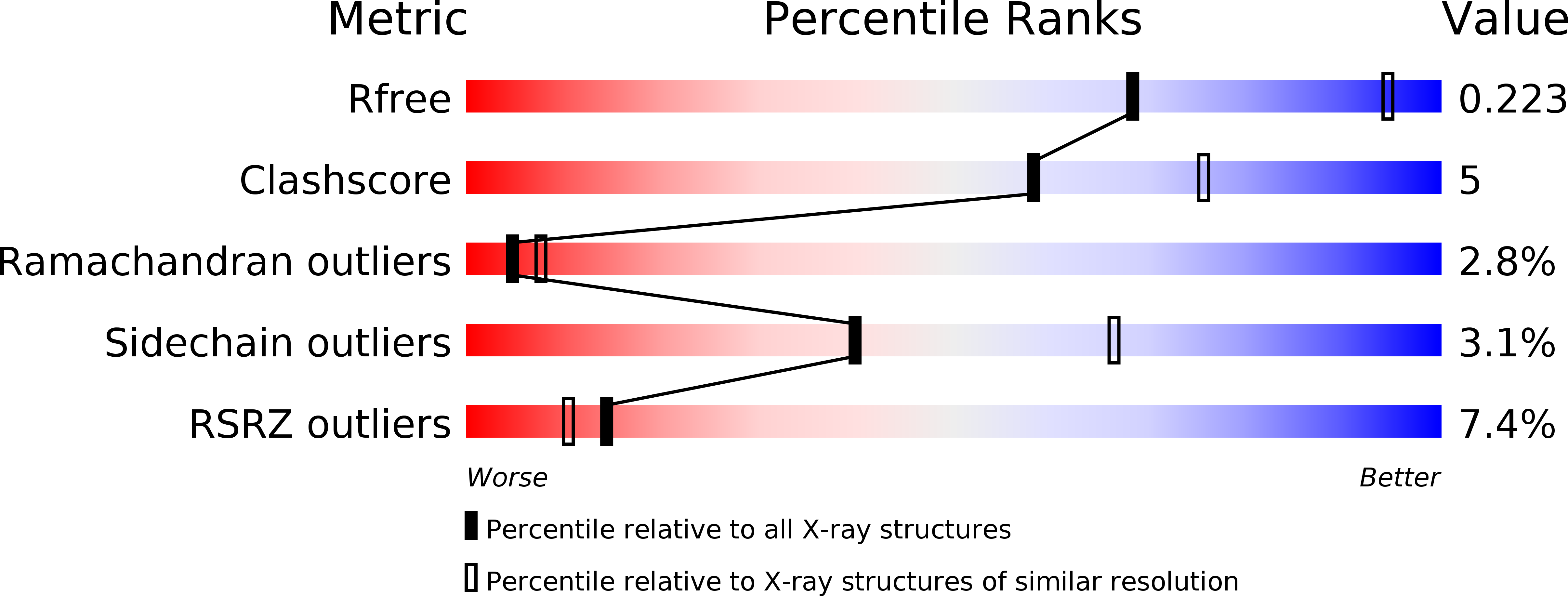
Resolution:
2.60 Å
R-Value Free:
0.28
R-Value Work:
0.22
R-Value Observed:
0.22
Space Group:
P 63
Macromolecular Entities
Structures with similar UniProt ID
Protein Blast
Polymer Type:polypeptide(L)
Molecule:Myosin A Tail Interacting Protein
Chain IDs:A, B, C
Chain Length:126
Number of Molecules:3
Biological Source:Plasmodium knowlesi
Structures with similar UniProt ID
Protein Blast
Polymer Type:polypeptide(L)
Molecule:Myosin A
Chain IDs:D
Chain Length:15
Number of Molecules:1
Biological Source:
Modified Residue
Compound ID Chain ID Parent Comp ID Details 2D Image
MSE A MET SELENOMETHIONINE
SAC D SER N-ACETYL-SERINE
Primary Citation
Structure of the MTIP-MyoA complex, a key component of the malaria parasite invasion motor.
Proc.Natl.Acad.Sci.Usa 103 4852 4857 (2006)
PMID: 16547135 DOI: 10.1073/pnas.0510907103

Abstact

The causative agents of malaria have developed a sophisticated machinery for entering multiple cell types in the human and insect hosts. In this machinery, a critical interaction occurs between the unusual myosin motor MyoA and the MyoA-tail Interacting Protein (MTIP). Here we present one crystal structure that shows three different conformations of Plasmodium MTIP, one of these in complex with the MyoA-tail, which reveal major conformational changes in the C-terminal domain of MTIP upon binding the MyoA-tail helix, thereby creating several hydrophobic pockets in MTIP that are the recipients of key hydrophobic side chains of MyoA. Because we also show that the MyoA helix is able to block parasite growth, this provides avenues for designing antimalarials.

Legend

Protein

Chemical

Disease

Primary Citation of related structures