2ATE image
Deposition Date 2005-08-24
Release Date 2006-08-29
Last Version Date 2024-11-13
Entry Detail
PDB ID:
2ATE
Keywords:
Title:
Structure of the complex of PurE with NitroAIR
Biological Source:
Source Organism:
Escherichia coli (Taxon ID: 562)
Host Organism:
Method Details:
Experimental Method:
Resolution:
1.80 Å
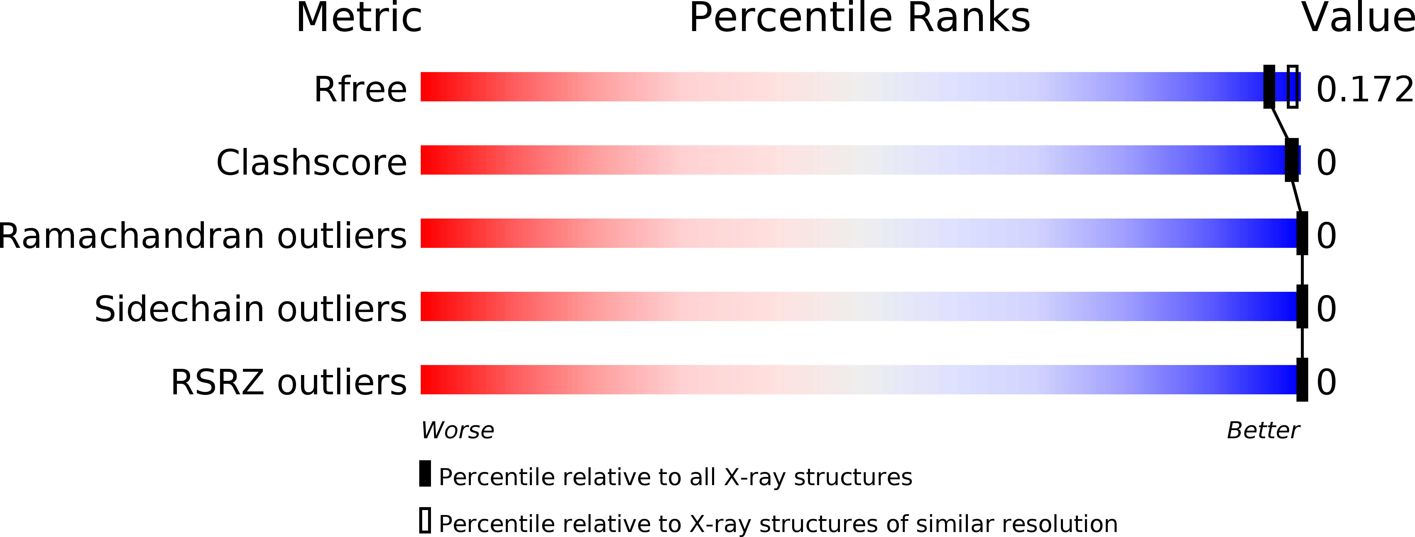
R-Value Free:
0.20
R-Value Work:
0.14
R-Value Observed:
0.15
Space Group:
I 4 2 2
Macromolecular Entities
Structures with similar UniProt ID
Protein Blast
Polymer Type:polypeptide(L)
Molecule:Phosphoribosylaminoimidazole carboxylase catalytic subunit
Gene (Uniprot):purE
Chain IDs:A
Chain Length:169
Number of Molecules:1
Biological Source:Escherichia coli
Modified Residue
Compound ID Chain ID Parent Comp ID Details 2D Image
MSE A MET SELENOMETHIONINE
Ligand Molecules
Primary Citation
N5-CAIR mutase: role of a CO2 binding site and substrate movement in catalysis.
Biochemistry 46 2842 2855 (2007)
PMID: 17298082 DOI: 10.1021/bi602436g

Abstact

N5-Carboxyaminoimidazole ribonucleotide mutase (N5-CAIR mutase or PurE) from Escherichia coli catalyzes the reversible interconversion of N5-CAIR to carboxyaminoimidazole ribonucleotide (CAIR) with direct CO2 transfer. Site-directed mutagenesis, a pH-rate profile, DFT calculations, and X-ray crystallography together provide new insight into the mechanism of this unusual transformation. These studies suggest that a conserved, protonated histidine (His45) plays an essential role in catalysis. The importance of proton transfers is supported by DFT calculations on CAIR and N5-CAIR analogues in which the ribose 5'-phosphate is replaced with a methyl group. The calculations suggest that the nonaromatic tautomer of CAIR (isoCAIR) is only 3.1 kcal/mol higher in energy than its aromatic counterpart, implicating this species as a potential intermediate in the PurE-catalyzed reaction. A structure of wild-type PurE cocrystallized with 4-nitroaminoimidazole ribonucleotide (NO2-AIR, a CAIR analogue) and structures of H45N and H45Q PurEs soaked with CAIR have been determined and provide the first insight into the binding of an intact PurE substrate. A comparison of 19 available structures of PurE and PurE mutants in apo and nucleotide-bound forms reveals a common, buried carboxylate or CO2 binding site for CAIR and N5-CAIR in a hydrophobic pocket in which the carboxylate or CO2 interacts with backbone amides. This work has led to a mechanistic proposal in which the carboxylate orients the substrate for proton transfer from His45 to N5-CAIR to form an enzyme-bound aminoimidazole ribonucleotide (AIR) and CO2 intermediate. Subsequent movement of the aminoimidazole moiety of AIR reorients it for addition of CO2 at C4 to generate isoCAIR. His45 is now in a position to remove a C4 proton to produce CAIR.

Legend

Protein

Chemical

Disease

Primary Citation of related structures