2ASQ image
Deposition Date 2005-08-23
Release Date 2005-10-11
Last Version Date 2024-05-22
Entry Detail
PDB ID:
2ASQ
Keywords:
Title:
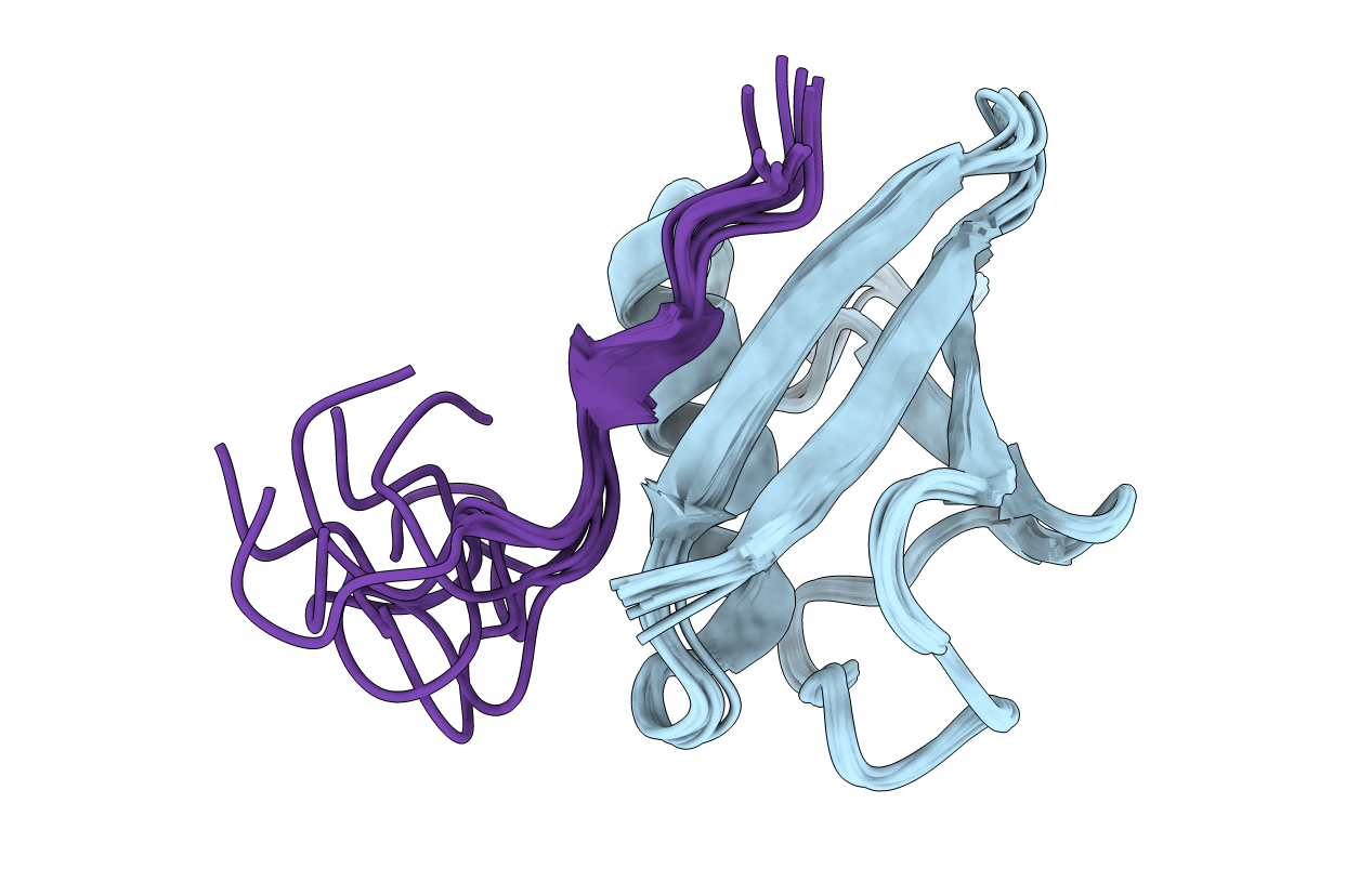
Solution Structure of SUMO-1 in Complex with a SUMO-binding Motif (SBM)
Biological Source:
Source Organism:
Homo sapiens (Taxon ID: 9606)
Host Organism:
Method Details:
Experimental Method:
Conformers Calculated:
10
Conformers Submitted:
10
Selection Criteria:
all calculated structures submitted
Macromolecular Entities
Polymer Type:polypeptide(L)
Molecule:Small ubiquitin-related modifier 1
Gene (Uniprot):SUMO1
Chain IDs:A
Chain Length:97
Number of Molecules:1
Biological Source:Homo sapiens
Polymer Type:polypeptide(L)
Molecule:Protein inhibitor of activated STAT2
Gene (Uniprot):PIAS2
Chain IDs:B
Chain Length:25
Number of Molecules:1
Biological Source:Homo sapiens
Ligand Molecules
Primary Citation
Small Ubiquitin-like Modifier (SUMO) Recognition of a SUMO Binding Motif: A reversal of the bound orientation
J.Biol.Chem. 280 40122 40129 (2005)
PMID: 16204249 DOI: 10.1074/jbc.M507059200

Abstact

Sumoylation has recently been identified as an important mechanism that regulates protein interactions and localization in essential cellular functions, such as gene transcription, subnuclear structure formation, viral infection, and cell cycle progression. A SUMO binding amino acid sequence motif (SBM), which recognizes the SUMO moiety of modified proteins in sumoylation-dependent cellular functions, has been consistently identified by several recent studies. To understand the mechanism of SUMO recognition by the SBM, we have solved the solution structure of SUMO-1 in complex with a peptide containing the SBM derived from the protein PIASX (KVDVIDLTIESSSDEEEDPPAKR). Surprisingly, the structure reveals that the bound orientation of the SBM can reverse depending on the sequence context. The structure also reveals a novel mechanism of recognizing target sequences by a ubiquitin-like module. Unlike ubiquitin binding motifs, which all form helices and bind to the main beta-sheet of ubiquitin, the SBM forms an extended structure that binds between the alpha-helix and a beta-strand of SUMO-1. This study provides a clear mechanism of the SBM sequence variations and its recognition of the SUMO moiety in sumoylated proteins.

Legend

Protein

Chemical

Disease

Primary Citation of related structures