2ARV image
Deposition Date 2005-08-22
Release Date 2006-03-07
Last Version Date 2024-10-30
Entry Detail
PDB ID:
2ARV
Title:
Structure of human Activin A
Biological Source:
Source Organism:
Homo sapiens (Taxon ID: 9606)
Method Details:
Experimental Method:
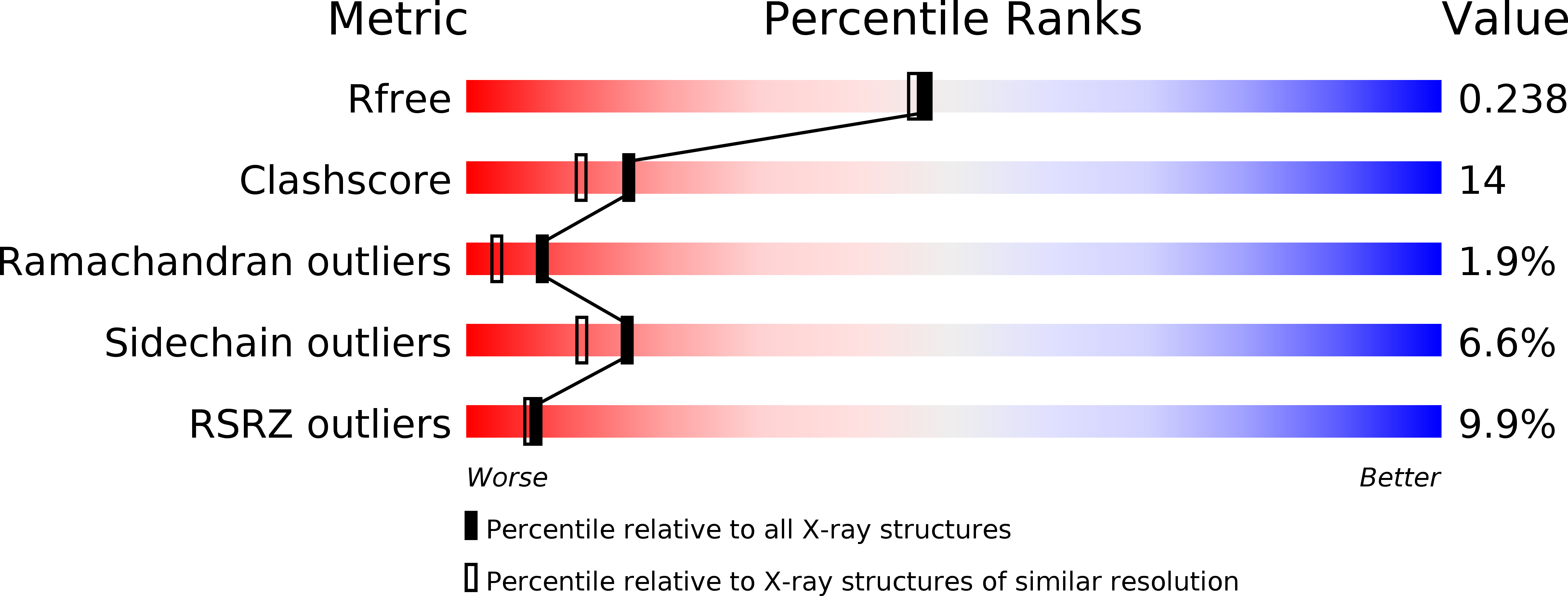
Resolution:
2.00 Å
R-Value Free:
0.25
R-Value Work:
0.21
R-Value Observed:
0.21
Space Group:
I 2 2 2
Macromolecular Entities
Structures with similar UniProt ID
Protein Blast
Polymer Type:polypeptide(L)
Molecule:Inhibin beta A chain
Gene (Uniprot):INHBA
Chain IDs:A, B
Chain Length:116
Number of Molecules:2
Biological Source:Homo sapiens
Primary Citation
Structural basis for the inhibition of activin signalling by follistatin
Embo J. 25 1035 1045 (2006)
PMID: 16482217 DOI: 10.1038/sj.emboj.7601000

Abstact

The secreted, multidomain protein follistatin binds activins with high affinity, inhibiting their receptor interaction. We have dissected follistatin's domain structure and shown that the minimal activin-inhibiting fragment of follistatin is comprised of the first and second Fs domains (Fs12). This protein can bind to activin dimer and form a stable complex containing two Fs12 molecules and one activin dimer. We have solved crystal structures of activin A alone and its complex with Fs12 fragment to 2 A resolution. The complex structure shows how Fs12 molecules wrap around the back of the 'wings' of activin, blocking the type II receptor-binding site on activin A. Arginine 192 in Fs2 is a key residue in this interaction, inserting itself in between activin's fingers. Complex formation imposes a novel orientation for the EGF- and Kazal-like subdomains in the Fs2 domain and activin A shows further variation from the canonical TGF-beta family fold. The structure provides a detailed description of the inhibitory mechanism and gives insights into interactions of follistatin with other TGF-beta family proteins.

Legend

Protein

Chemical

Disease

Primary Citation of related structures
Feedback Form
Name
Email
Institute
Feedback