Deposition Date
1996-10-30
Release Date
1997-05-15
Last Version Date
2024-05-22
Entry Detail
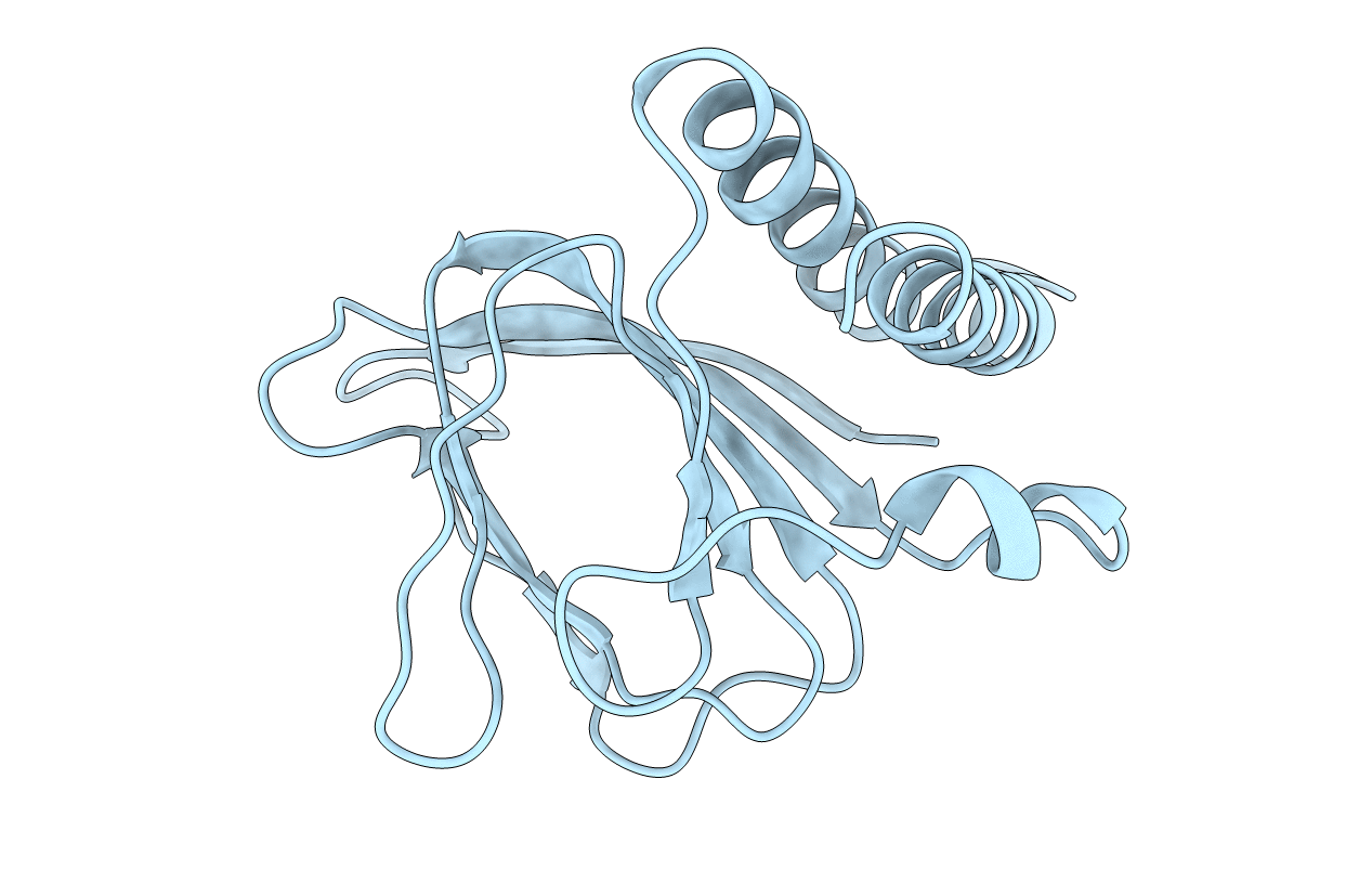
PDB ID:
2ARA
Keywords:
Title:
APO FORM OF ESCHERICHIA COLI REGULATORY PROTEIN ARAC
Biological Source:
Source Organism(s):
Escherichia coli (Taxon ID: 562)
Expression System(s):
Method Details:
Experimental Method:
Resolution:
2.80 Å
R-Value Free:
0.33
R-Value Work:
0.21
R-Value Observed:
0.21
Space Group:
P 31 2 1