2AQX image
Deposition Date 2005-08-18
Release Date 2005-12-06
Last Version Date 2024-03-13
Entry Detail
PDB ID:
2AQX
Keywords:
Title:
Crystal Structure of the Catalytic and CaM-Binding domains of Inositol 1,4,5-Trisphosphate 3-Kinase B
Biological Source:
Source Organism:
Mus musculus (Taxon ID: 10090)
Host Organism:
Method Details:
Experimental Method:
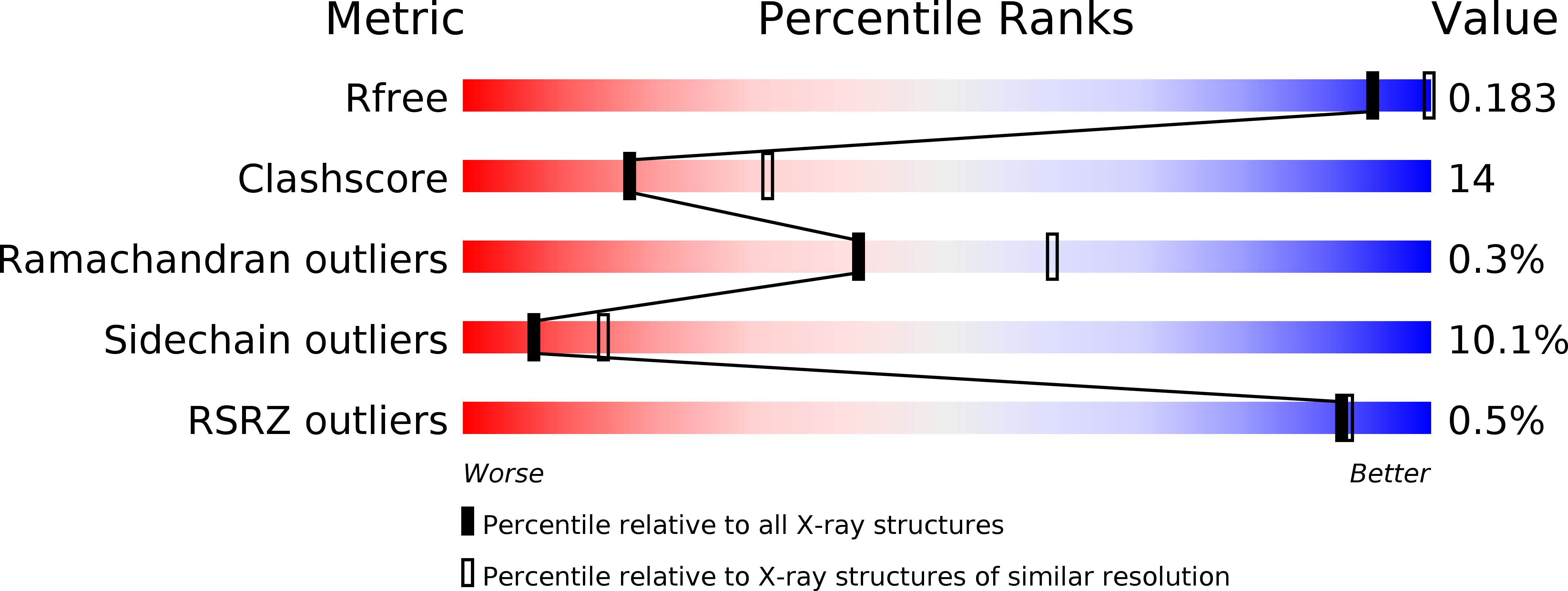
Resolution:
2.50 Å
R-Value Free:
0.26
R-Value Work:
0.18
R-Value Observed:
0.18
Space Group:
P 1
Macromolecular Entities
Structures with similar UniProt ID
Protein Blast
Polymer Type:polypeptide(L)
Molecule:PREDICTED: inositol 1,4,5-trisphosphate 3-kinase B
Chain IDs:A, B
Chain Length:289
Number of Molecules:2
Biological Source:Mus musculus
Primary Citation
Structural insights into enzyme regulation for inositol 1,4,5-trisphosphate 3-kinase B
Biochemistry 44 14486 14493 (2005)
PMID: 16262249 DOI: 10.1021/bi051256q

Abstact

D-Myoinositol 1,4,5-trisphophate 3-kinases (IP(3)-3Ks) play important roles in metazoan cellular signaling. It has been demonstrated that mice without a functional version of IP(3)-3K isoform B are deficient in peripheral T-cells, indicating that IP(3)-3KB is essential to the developing immune system. The recent apo IP(3)-3KA structure exhibited a helix at the catalytic domain N-terminus exhibited a helix at the N-terminus of the catalytic domain, with a tryptophan indole moiety mimicking the binding mode of the substrate ATP purine ring, suggesting a mechanism of autoinhibition. Here we present the structure of the complete catalytic domain of IP(3)-3KB, including the CaM binding domain in complex with Mg(2+) and ATP. The crystal structure reveals a homodimeric arrangement of IP(3)-3KB catalytic domains, mediated via an intermolecular antiparallel beta-sheet formed from part of the CaM binding region. Residues from the putative autoinhibitory helix are rearranged into a loop configuration, with extensive interactions with the bound ATP. Mutagenesis of residues from this region reveals that substitution of the putative autoinhibitory tryptophan generates a hyperactive enzyme which retains Ca(2+)/CaM sensitivity. The IP(3)-3KB structure suggests a mechanism of enzyme activation, and raises the possibility that an interaction between IP(3)-3KB molecules may occur as part of the catalytic or regulatory cycle.

Legend

Protein

Chemical

Disease

Primary Citation of related structures
Feedback Form
Name
Email
Institute
Feedback