Deposition Date
2005-08-18
Release Date
2006-05-23
Last Version Date
2024-05-29
Entry Detail
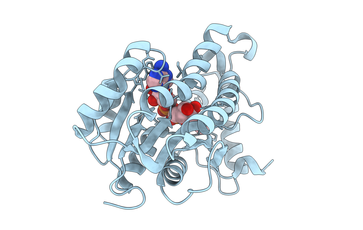
PDB ID:
2AQK
Keywords:
Title:
Crystal structure of Isoniazid-resistant S94A Enoyl-ACP(CoA) reductase mutant enzyme from Mycobacterium tuberculosis in complex with NADH
Biological Source:
Source Organism(s):
Mycobacterium tuberculosis (Taxon ID: 1773)
Expression System(s):
Method Details:
Experimental Method:
Resolution:
2.30 Å
R-Value Free:
0.24
R-Value Work:
0.18
R-Value Observed:
0.19
Space Group:
P 62 2 2