Deposition Date
2005-08-11
Release Date
2006-07-25
Last Version Date
2024-02-14
Entry Detail
PDB ID:
2ANQ
Keywords:
Title:
Crystal Structure of E.coli DHFR in complex with NADPH and the inhibitor compound 10a.
Biological Source:
Source Organism(s):
Escherichia coli (Taxon ID: 562)
Expression System(s):
Method Details:
Experimental Method:
Resolution:
2.13 Å
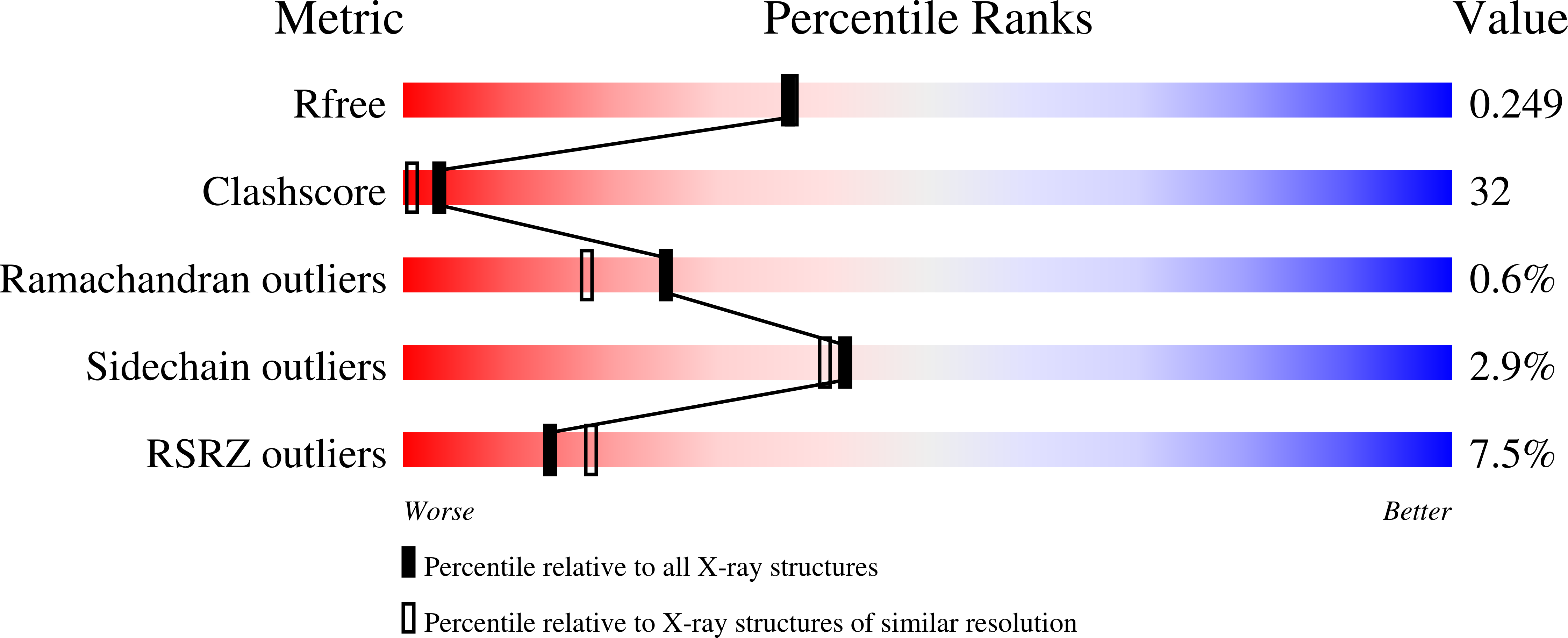
R-Value Free:
0.25
R-Value Work:
0.23
R-Value Observed:
0.23
Space Group:
P 21 21 21