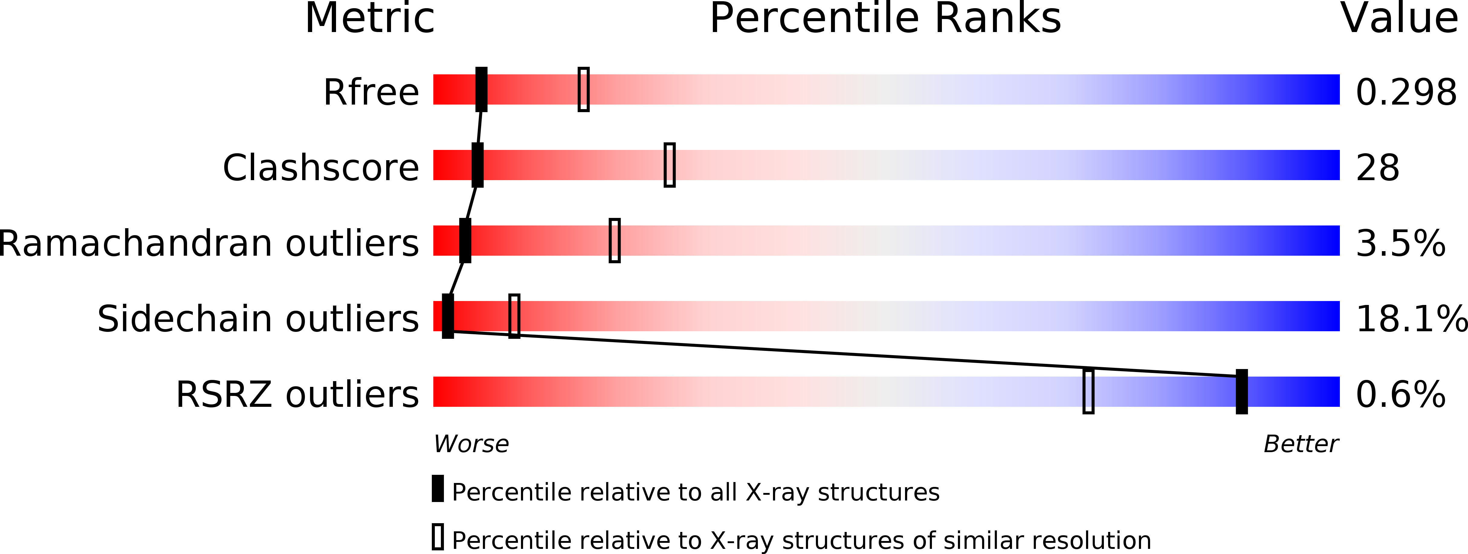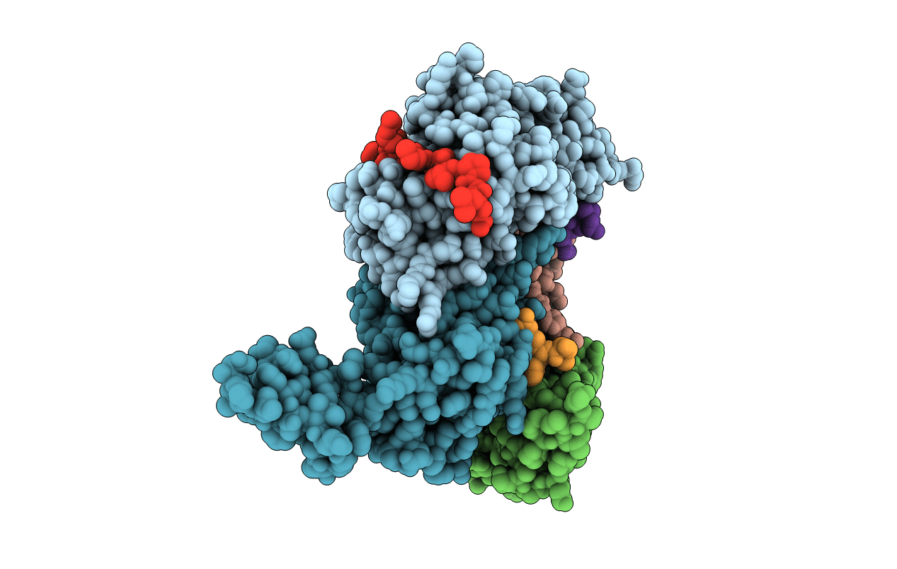
2AN6 image
Deposition Date 2005-08-11
Release Date 2006-05-30
Last Version Date 2024-03-13
Entry Detail
PDB ID:
2AN6
Keywords:
Title:
Protein-peptide complex
Biological Source:
Source Organism:
Mus musculus (Taxon ID: 10090)
Host Organism:
Method Details:
Experimental Method:
Resolution:
3.00 Å
R-Value Free:
0.30
R-Value Work:
0.23
R-Value Observed:
0.24
Space Group:
P 1 21 1
Macromolecular Entities
Structures with similar UniProt ID
Protein Blast
Polymer Type:polypeptide(L)
Molecule:Ubiquitin ligase SIAH1A
Gene (Uniprot):Siah1a
Chain IDs:A, B, C, D
Chain Length:191
Number of Molecules:4
Biological Source:Mus musculus
Polymer Type:polypeptide(L)
Molecule:peptide from Phyllopod
Chain IDs:E, F, G, H
Chain Length:24
Number of Molecules:4
Biological Source:
Ligand Molecules
Primary Citation
Elucidation of the substrate binding site of Siah ubiquitin ligase
Structure 14 695 701 (2006)
PMID: 16615911 DOI: 10.1016/j.str.2005.12.013

Abstact

The Siah family of RING proteins function as ubiquitin ligase components, contributing to the degradation of multiple targets involved in cell growth, differentiation, angiogenesis, oncogenesis, and inflammation. Previously, a binding motif (degron) was recognized in many of the Siah degradation targets, suggesting that Siah itself may facilitate substrate recognition. We report the crystal structure of the Siah in complex with a peptide containing the degron motif. Binding is within a groove formed in part by the zinc fingers and the first two beta strands of the TRAF-C domain of Siah. We show that residues in the degron, previously described to facilitate binding to Siah, interact with the protein. Mutagenesis of Siah at sites of interaction also abrogates both in vitro peptide binding and destabilization of a known Siah target.

Legend

Protein

Chemical

Disease

Primary Citation of related structures