Deposition Date
2005-08-05
Release Date
2006-01-17
Last Version Date
2024-11-06
Entry Detail
PDB ID:
2ALA
Keywords:
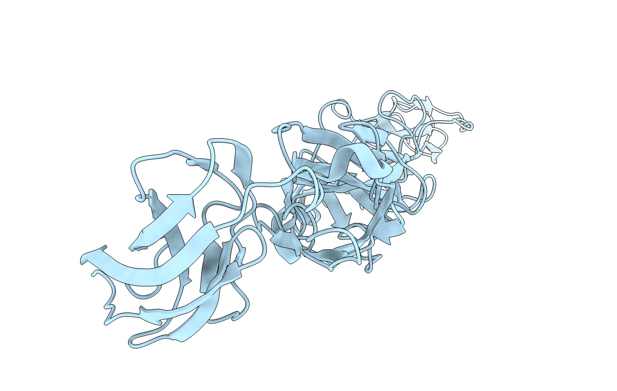
Title:
Crystal structure of the Semliki Forest Virus envelope protein E1 in its monomeric conformation.
Biological Source:
Source Organism(s):
Semliki forest virus (Taxon ID: 11033)
Method Details:
Experimental Method:
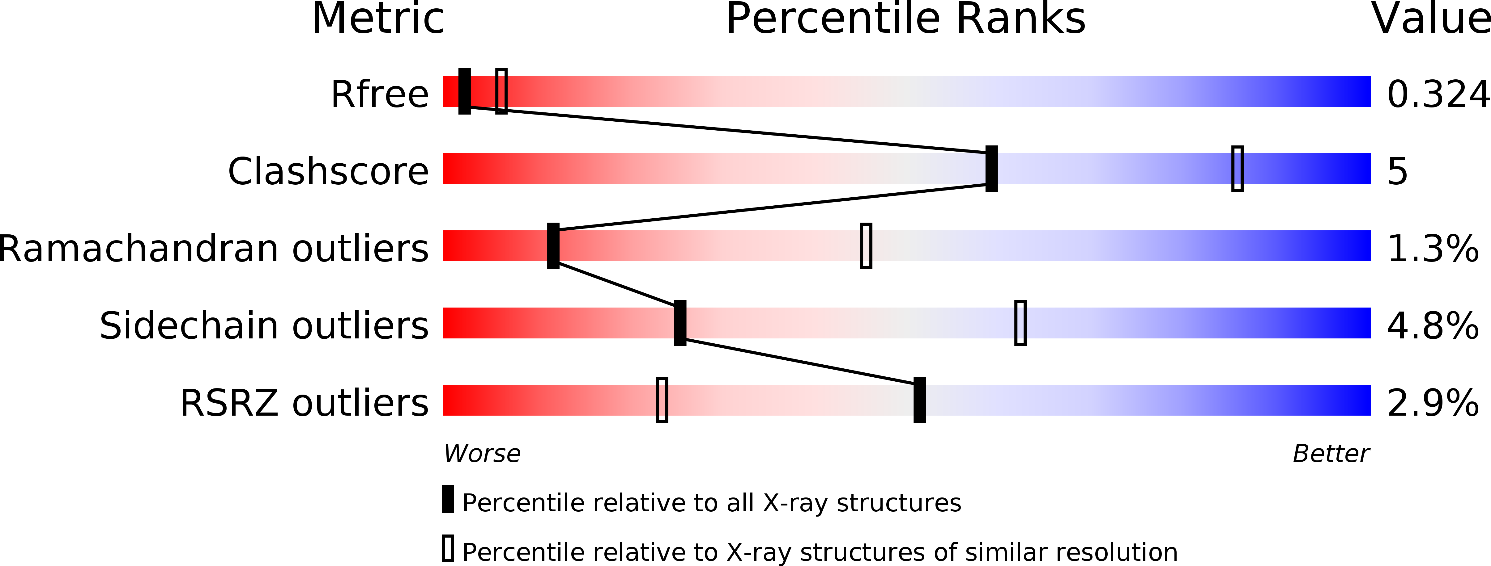
Resolution:
3.00 Å
R-Value Free:
0.31
R-Value Work:
0.26
R-Value Observed:
0.27
Space Group:
P 64 2 2