2AKQ image
Deposition Date 2005-08-03
Release Date 2005-08-16
Last Version Date 2024-10-23
Entry Detail
PDB ID:
2AKQ
Title:
The structure of bovine B-lactoglobulin A in crystals grown at very low ionic strength
Biological Source:
Source Organism:
Bos taurus (Taxon ID: 9913)
Method Details:
Experimental Method:
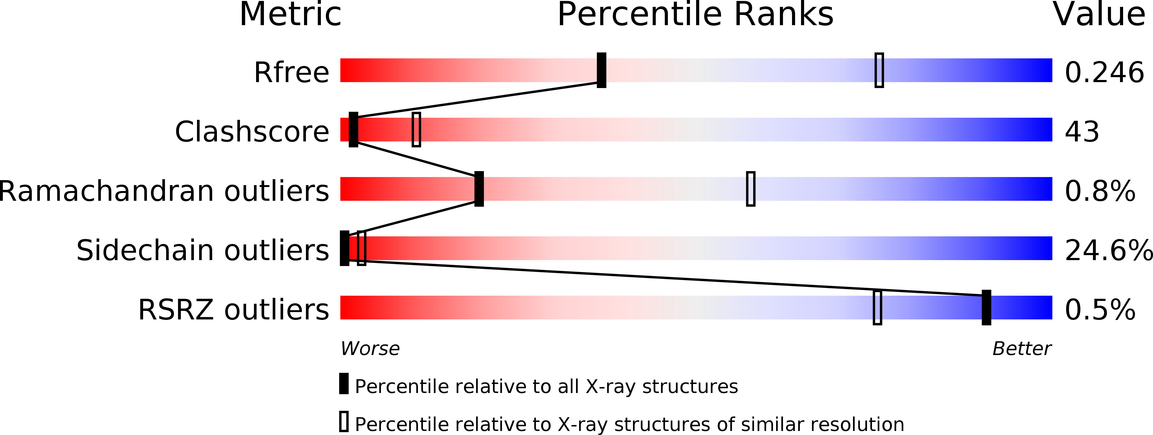
Resolution:
3.00 Å
R-Value Free:
0.26
R-Value Work:
0.22
R-Value Observed:
0.21
Space Group:
P 21 21 21
Macromolecular Entities
Polymer Type:polypeptide(L)
Molecule:Beta-lactoglobulin variant A
Gene (Uniprot):LGB
Chain IDs:A, B, C, D
Chain Length:162
Number of Molecules:4
Biological Source:Bos taurus
Primary Citation

Abstact

Bovine beta-lactoglobulin (BLG) is a globular protein of uncertain physiological function and a member of the lipocalin superfamily of proteins. Here, we present the X-ray structure at 3.0 angstroms of BLG (variant A) from an orthorhombic (P2(1)2(1)2(1)) pseudo-tetragonal crystal form that suffers from pseudo-merohedral twinning (final R(working) = 0.224, R(free) = 0.265). Crystals were grown by dialysis against ultra-purified water (i.e., at very low ionic strength), at pH approximately 5.2 (approximately pI), conditions vastly different from all other BLG structures determined previously. This allows critical assessment of the BLG structure and of the influence that pH, ionic strength, and crystal packing may have on the molecular structure of BLG. The pH-sensitive EF loop is found in the closed conformation characteristic of BLG at pH less than 7 and moderate to high ionic strength. Although the hydrophobic pocket appears to be empty, the presence of highly disordered water molecules cannot be excluded. The dimer interface and the hydrophobic pocket (calyx) are preserved. However, the orientation of the subunits in the dimer varies considerably with crystal form. Structure is deposited with PDB ID 2akq.

Legend

Protein

Chemical

Disease

Primary Citation of related structures