2AK3 image
Deposition Date 1995-03-07
Release Date 1995-05-08
Last Version Date 2024-02-14
Entry Detail
PDB ID:
2AK3
Title:
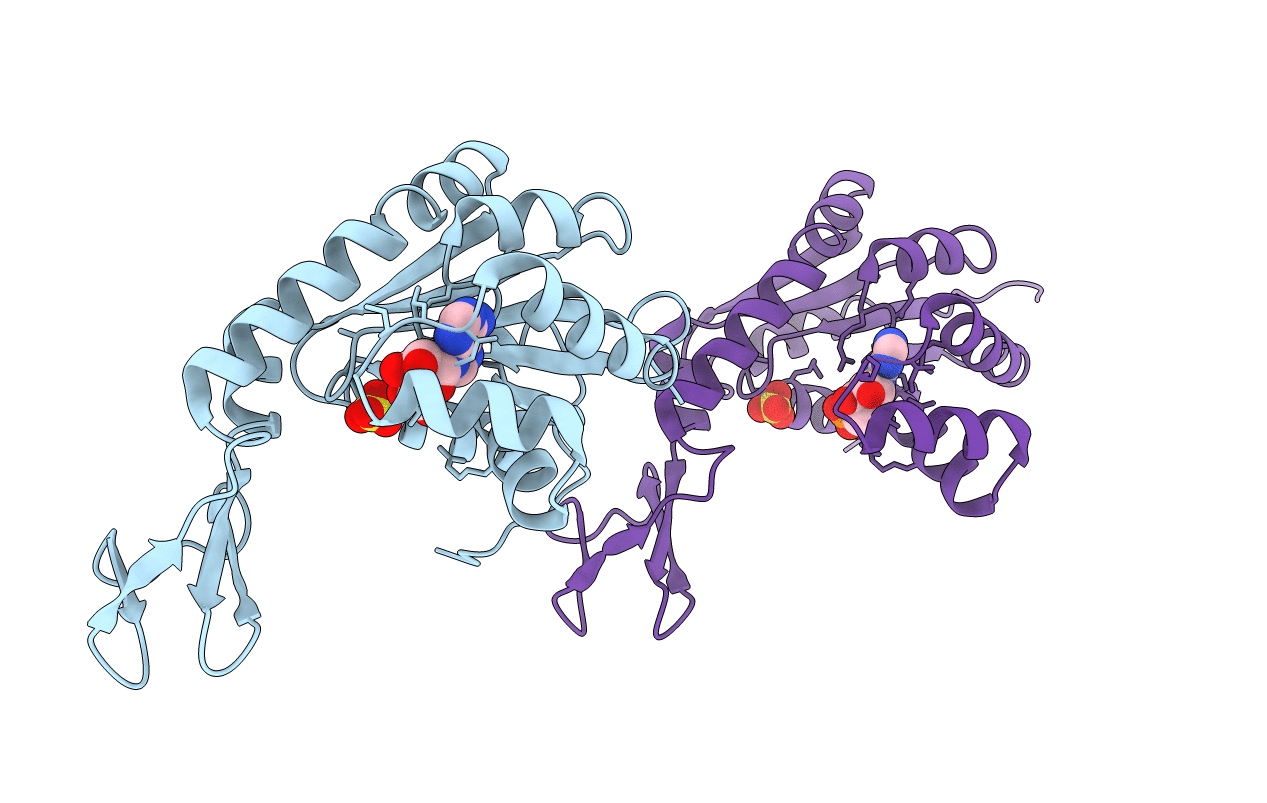
THE THREE-DIMENSIONAL STRUCTURE OF THE COMPLEX BETWEEN MITOCHONDRIAL MATRIX ADENYLATE KINASE AND ITS SUBSTRATE AMP AT 1.85 ANGSTROMS RESOLUTION
Biological Source:
Source Organism:
Bos taurus (Taxon ID: 9913)
Method Details:
Experimental Method:
Resolution:
1.85 Å
R-Value Work:
0.18
R-Value Observed:
0.18
Space Group:
P 21 21 21
Macromolecular Entities
Polymer Type:polypeptide(L)
Molecule:ADENYLATE KINASE ISOENZYME-3
Gene (Uniprot):AK3
Chain IDs:A, B
Chain Length:226
Number of Molecules:2
Biological Source:Bos taurus
Primary Citation
The refined structure of the complex between adenylate kinase from beef heart mitochondrial matrix and its substrate AMP at 1.85 A resolution.
J.Mol.Biol. 217 541 549 (1991)
PMID: 1994037 DOI: 10.1016/0022-2836(91)90756-V

Abstact

The crystal structure of the complex between adenylate kinase from bovine mitochondrial matrix and its substrate AMP has been refined at 1.85 A resolution (1 A = 0.1 nm). Based on 42,519 independent reflections of better than 10 A resolution, a final R-factor of 18.9% was obtained with a model obeying standard geometry within 0.016 A in bond lengths and 3.2 degrees in bond angles. There are two enzyme: substrate complexes in the asymmetric unit, each consisting of 226 amino acid residues, one AMP and one sulfate ion. A superposition of the two full-length polypeptides revealed deviations that can be described as small relative movements of three domains. Best superpositions of individual domains yielded a residual overall root-mean-square deviation of 0.3 A for the backbone atoms and 0.5 A for the sidechains. The final model contains 381 solvent molecules in the asymmetric unit, 2 x 72 = 144 of which occupy corresponding positions in both complexes.

Legend

Protein

Chemical

Disease

Primary Citation of related structures