Deposition Date
2005-08-01
Release Date
2005-09-20
Last Version Date
2024-12-25
Entry Detail
PDB ID:
2AJF
Keywords:
Title:
Structure of SARS coronavirus spike receptor-binding domain complexed with its receptor
Biological Source:
Source Organism(s):
Homo sapiens (Taxon ID: 9606)
SARS coronavirus (Taxon ID: 227859)
SARS coronavirus (Taxon ID: 227859)
Expression System(s):
Method Details:
Experimental Method:
Resolution:
2.90 Å
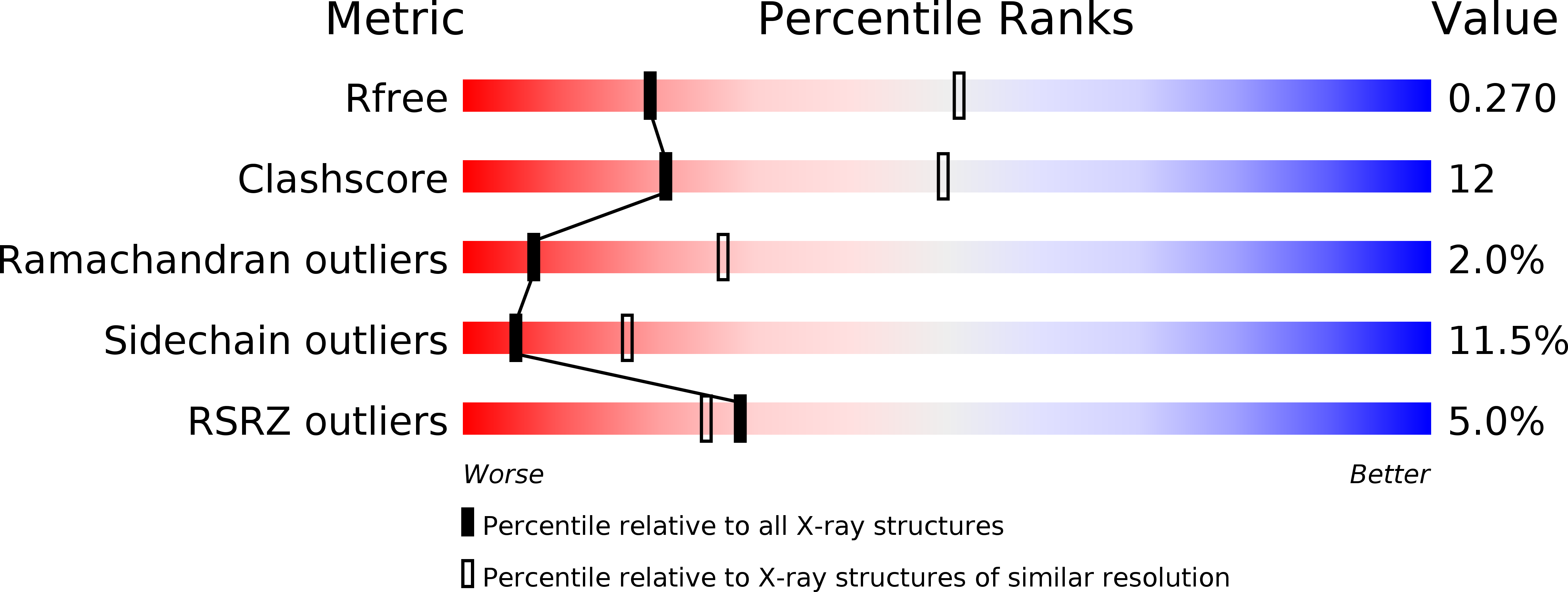
R-Value Free:
0.27
R-Value Work:
0.21
R-Value Observed:
0.22
Space Group:
P 1 21 1