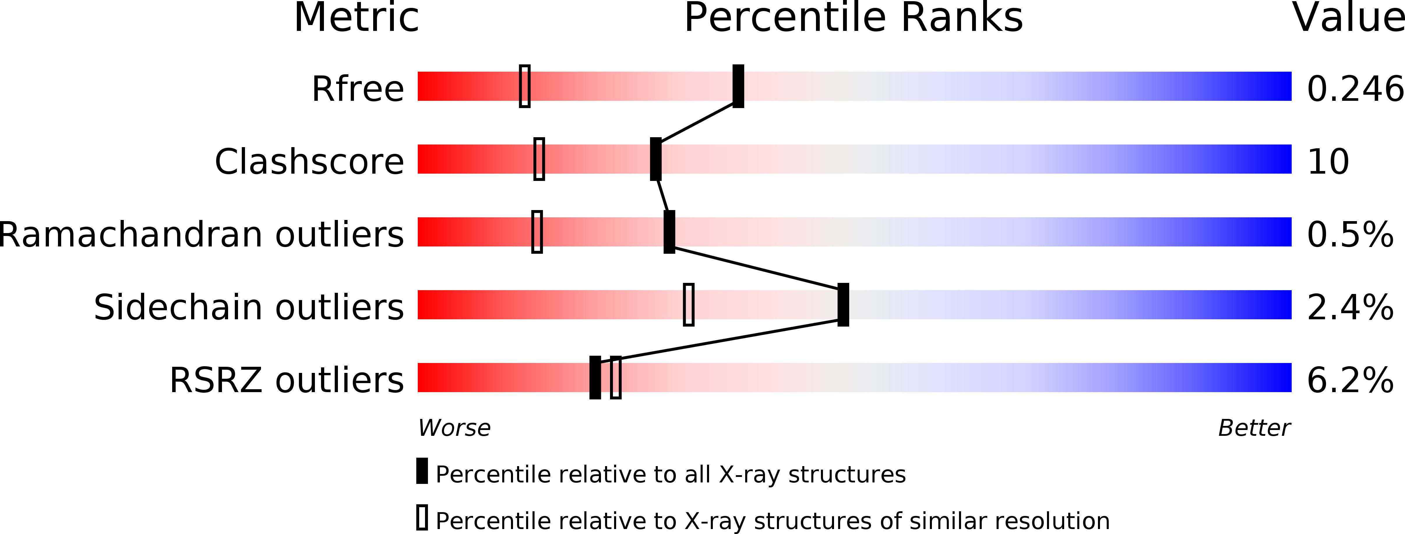
2AIE image
Entry Detail
PDB ID:
2AIE
Keywords:
Title:
S.pneumoniae polypeptide deformylase complexed with SB-505684
Biological Source:
Source Organism:
Host Organism:
PDB Version:
Deposition Date:
2005-07-29
Release Date:
2005-09-06
Method Details:
Experimental Method:
Resolution:
1.70 Å
R-Value Free:
0.25
R-Value Work:
0.22
Space Group:
P 43
Macromolecular Entities
Polymer Type:polypeptide(L)
Description:Peptide deformylase
Chain IDs:A (auth: P)
Chain Length:203
Number of Molecules:1
Biological Source:Streptococcus pneumoniae
Primary Citation
Structural Variation and inhibitor binding in polypeptide deformylase from four different bacterial species
Protein Sci. 12 349 360 (2003)
PMID: 12538898 DOI: 10.1110/ps.0229303

Abstact

Polypeptide deformylase (PDF) catalyzes the deformylation of polypeptide chains in bacteria. It is essential for bacterial cell viability and is a potential antibacterial drug target. Here, we report the crystal structures of polypeptide deformylase from four different species of bacteria: Streptococcus pneumoniae, Staphylococcus aureus, Haemophilus influenzae, and Escherichia coli. Comparison of these four structures reveals significant overall differences between the two Gram-negative species (E. coli and H. influenzae) and the two Gram-positive species (S. pneumoniae and S. aureus). Despite these differences and low overall sequence identity, the S1' pocket of PDF is well conserved among the four enzymes studied. We also describe the binding of nonpeptidic inhibitor molecules SB-485345, SB-543668, and SB-505684 to both S. pneumoniae and E. coli PDF. Comparison of these structures shows similar binding interactions with both Gram-negative and Gram-positive species. Understanding the similarities and subtle differences in active site structure between species will help to design broad-spectrum polypeptide deformylase inhibitor molecules.

Legend

Protein

Chemical

Disease

Primary Citation of related structures