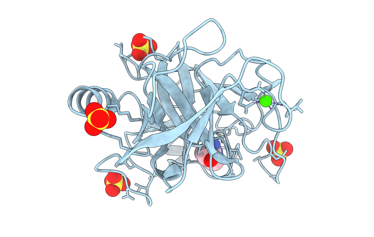
2AH4 image
Entry Detail
PDB ID:
2AH4
Keywords:
Title:
guanidinobenzoyl-trypsin acyl-enzyme at 1.13 A resolution
Biological Source:
Source Organism:
PDB Version:
Deposition Date:
2005-07-26
Release Date:
2006-05-16
Method Details:
Experimental Method:
Resolution:
1.13 Å
R-Value Free:
0.14
R-Value Work:
0.11
R-Value Observed:
0.12
Space Group:
P 21 21 21
Macromolecular Entities
Polymer Type:polypeptide(L)
Description:beta-trypsin
Chain IDs:A (auth: X)
Chain Length:223
Number of Molecules:1
Biological Source:Bos taurus
Primary Citation
Insights into the serine protease mechanism from atomic resolution structures of trypsin reaction intermediates
Proc.Natl.Acad.Sci.USA 103 6835 6840 (2006)
PMID: 16636277 DOI: 10.1073/pnas.0601910103

Abstact

Atomic resolution structures of trypsin acyl-enzymes and a tetrahedral intermediate analog, along with previously solved structures representing the Michaelis complex, are used to reconstruct events in the catalytic cycle of this classic serine protease. Structural comparisons provide insight into active site adjustments involved in catalysis. Subtle motions of the catalytic serine and histidine residues coordinated with translation of the substrate reaction center are seen to favor the forward progress of the acylation reaction. The structures also clarify the attack trajectory of the hydrolytic water in the deacylation reaction.

Legend

Protein

Chemical

Disease

Primary Citation of related structures