Deposition Date
2005-07-26
Release Date
2005-11-22
Last Version Date
2024-05-01
Entry Detail
PDB ID:
2AGH
Keywords:
Title:
Structural basis for cooperative transcription factor binding to the CBP coactivator
Biological Source:
Source Organism(s):
Mus musculus (Taxon ID: 10090)
Homo sapiens (Taxon ID: 9606)
Homo sapiens (Taxon ID: 9606)
Expression System(s):
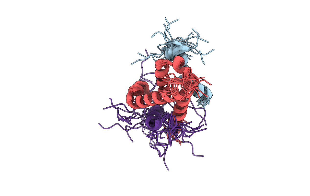
Method Details:
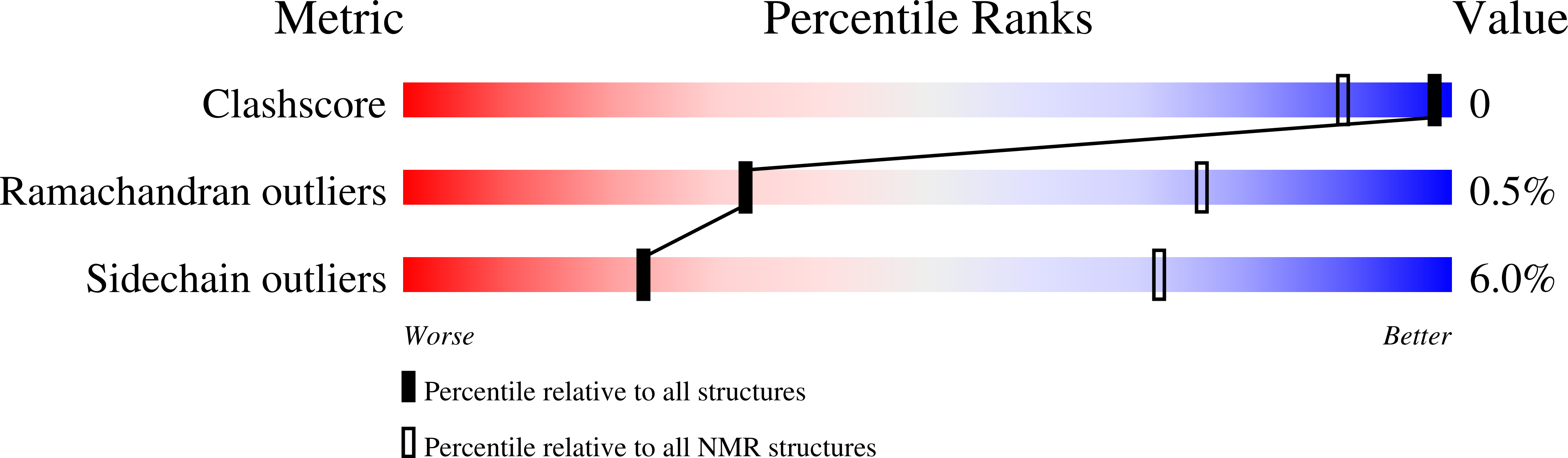
Experimental Method:
Conformers Calculated:
100
Conformers Submitted:
20
Selection Criteria:
lowest energy and lowest distance and angle violations