
Deposition Date
2005-07-21
Release Date
2006-03-28
Last Version Date
2025-03-26
Entry Detail
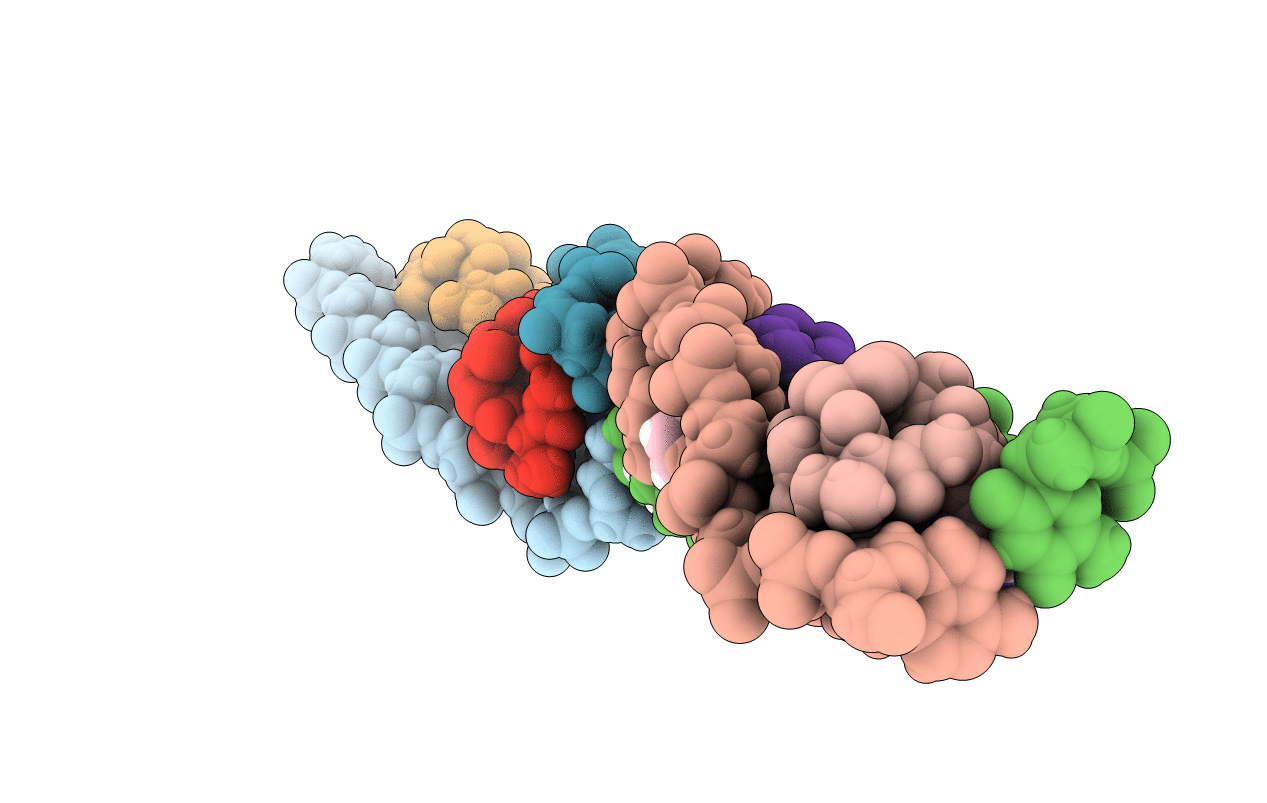
PDB ID:
2ADW
Keywords:
Title:
Crystal structure of Echinomycin-(ACGTACGT)2 solved by SAD
Biological Source:
Source Organism(s):
STREPTOMYCES ECHINATUS (Taxon ID: 67293)
Method Details:
Experimental Method:
Resolution:
1.60 Å
R-Value Free:
0.22
R-Value Observed:
0.18
Space Group:
P 42 21 2
