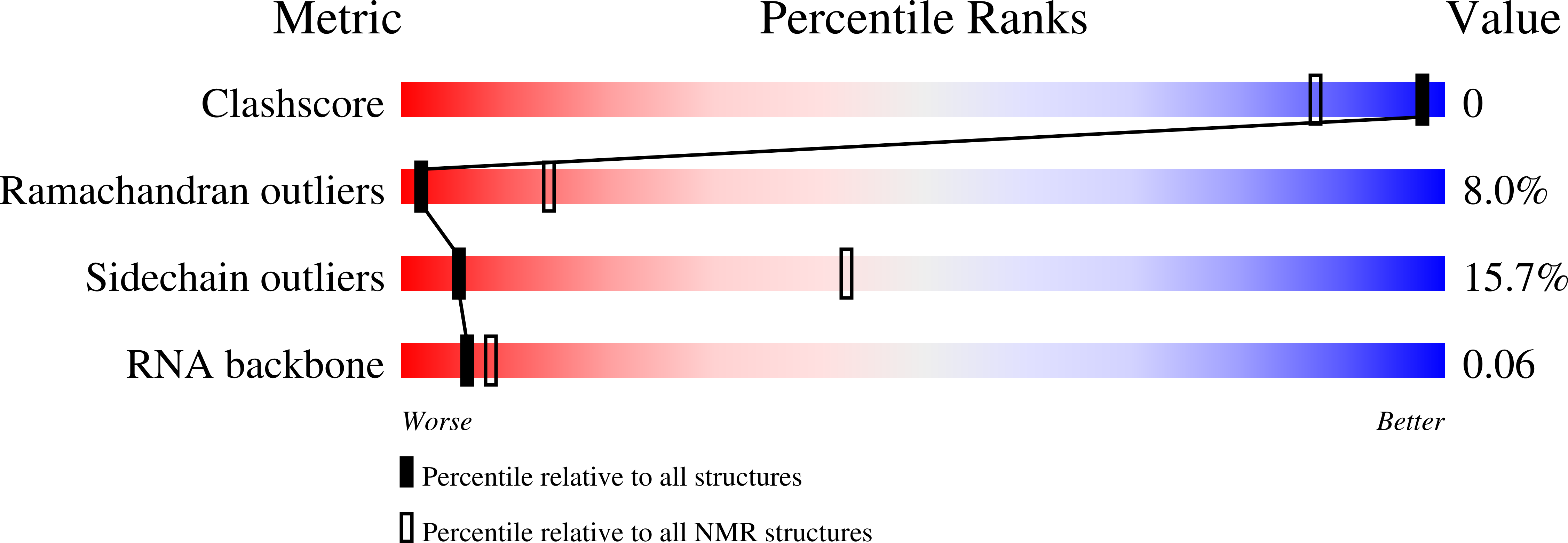
2ADB image
Deposition Date 2005-07-20
Release Date 2005-10-04
Last Version Date 2024-05-29
Entry Detail
PDB ID:
2ADB
Title:
Solution structure of Polypyrimidine Tract Binding protein RBD2 complexed with CUCUCU RNA
Biological Source:
Source Organism:
Homo sapiens (Taxon ID: 9606)
(Taxon ID: )
Host Organism:
Method Details:
Experimental Method:
Conformers Calculated:
40
Conformers Submitted:
20
Selection Criteria:
structures with the lowest energy
Macromolecular Entities
Polymer Type:polypeptide(L)
Molecule:Polypyrimidine tract-binding protein 1
Gene (Uniprot):PTBP1
Chain IDs:B (auth: A)
Chain Length:148
Number of Molecules:1
Biological Source:Homo sapiens
Polymer Type:polyribonucleotide
Molecule:5'-R(*CP*UP*CP*UP*CP*U)-3'
Chain IDs:A (auth: B)
Chain Length:6
Number of Molecules:1
Biological Source:
Ligand Molecules
Primary Citation
Structure of PTB bound to RNA: specific binding and implications for splicing regulation
Science 309 2054 2057 (2005)
PMID: 16179478 DOI: 10.1126/science.1114066

Abstact

The polypyrimidine tract binding protein (PTB) is a 58-kilodalton RNA binding protein involved in multiple aspects of messenger RNA metabolism, including the repression of alternative exons. We have determined the solution structures of the four RNA binding domains (RBDs) of PTB, each bound to a CUCUCU oligonucleotide. Each RBD binds RNA with a different binding specificity. RBD3 and RBD4 interact, resulting in an antiparallel orientation of their bound RNAs. Thus, PTB will induce RNA looping when bound to two separated pyrimidine tracts within the same RNA. This leads to structural models for how PTB functions as an alternative-splicing repressor.

Legend

Protein

Chemical

Disease

Primary Citation of related structures