
Deposition Date
2005-07-18
Release Date
2006-08-29
Last Version Date
2024-10-30
Entry Detail
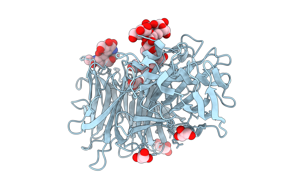
PDB ID:
2AC1
Keywords:
Title:
Crystal structure of a cell-wall invertase from Arabidopsis thaliana
Biological Source:
Source Organism(s):
Arabidopsis thaliana (Taxon ID: 3702)
Expression System(s):
Method Details:
Experimental Method:
Resolution:
2.15 Å
R-Value Free:
0.24
R-Value Work:
0.20
R-Value Observed:
0.20
Space Group:
C 2 2 21
