Deposition Date
2005-07-18
Release Date
2006-01-31
Last Version Date
2024-10-30
Entry Detail
PDB ID:
2ABZ
Keywords:
Title:
Crystal structure of C19A/C43A mutant of leech carboxypeptidase inhibitor in complex with bovine carboxypeptidase A
Biological Source:
Source Organism(s):
Bos taurus (Taxon ID: 9913)
Hirudo medicinalis (Taxon ID: 6421)
Hirudo medicinalis (Taxon ID: 6421)
Expression System(s):
Method Details:
Experimental Method:
Resolution:
2.16 Å
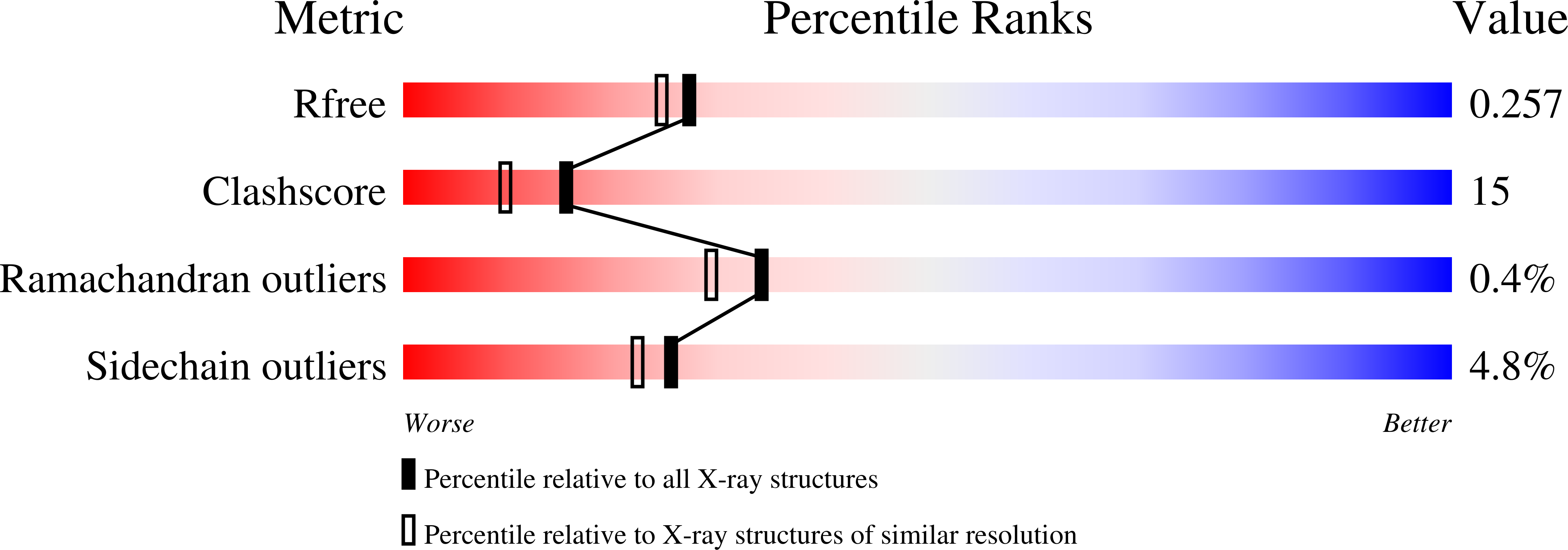
R-Value Free:
0.23
R-Value Work:
0.18
R-Value Observed:
0.19
Space Group:
P 43 21 2