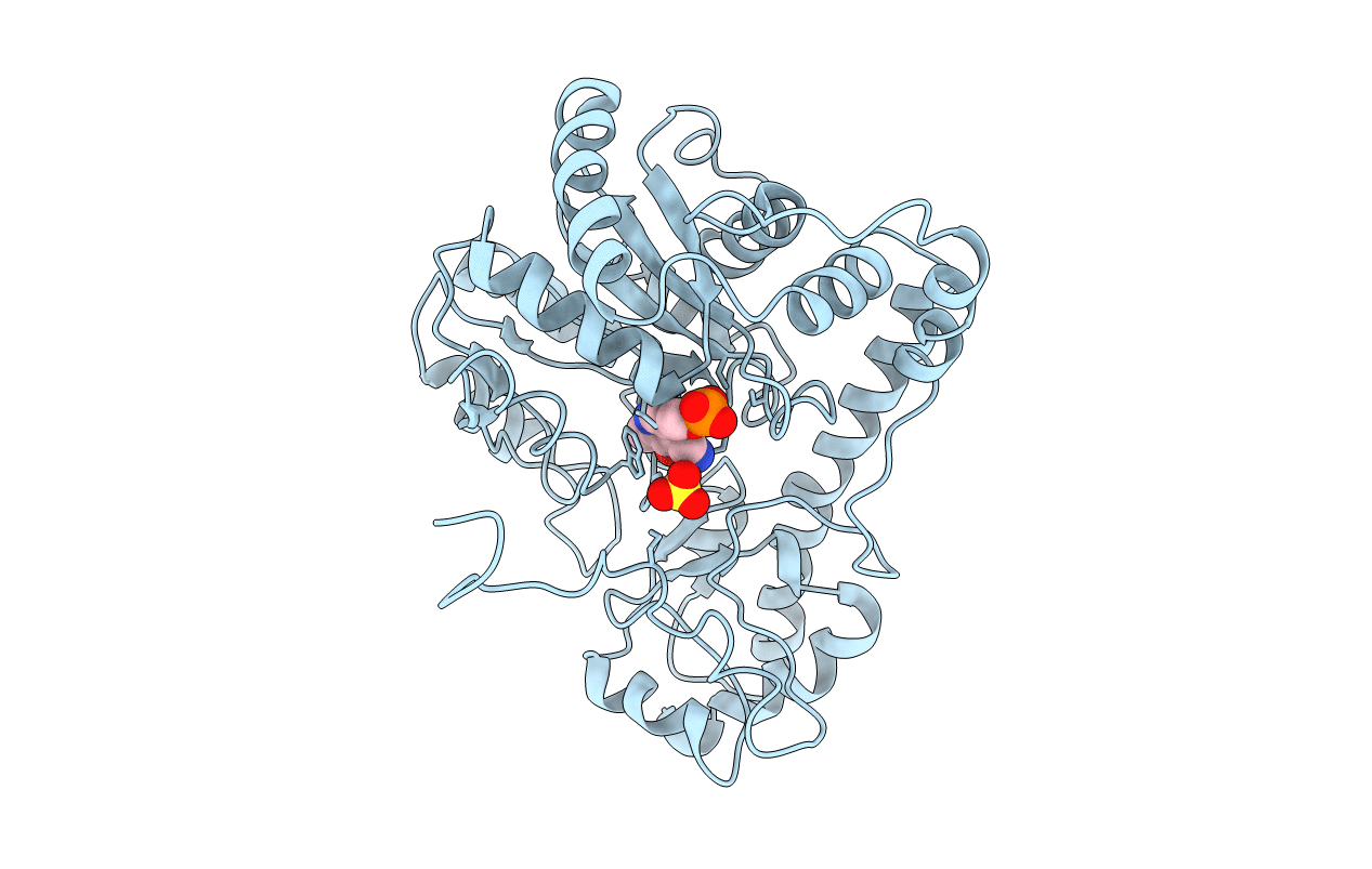
2AAT image
Entry Detail
PDB ID:
2AAT
Title:
2.8-ANGSTROMS-RESOLUTION CRYSTAL STRUCTURE OF AN ACTIVE-SITE MUTANT OF ASPARTATE AMINOTRANSFERASE FROM ESCHERICHIA COLI
Biological Source:
Source Organism:
Host Organism:
PDB Version:
Deposition Date:
1989-05-30
Release Date:
1989-10-15
Method Details:
Experimental Method:
Resolution:
2.80 Å
R-Value Observed:
0.22
Space Group:
C 2 2 21
Macromolecular Entities
Polymer Type:polypeptide(L)
Description:ASPARTATE AMINOTRANSFERASE
Mutations:K246A
Chain IDs:A
Chain Length:396
Number of Molecules:1
Biological Source:Escherichia coli
Primary Citation
2.8-A-resolution crystal structure of an active-site mutant of aspartate aminotransferase from Escherichia coli.
Biochemistry 28 8161 8167 (1989)
PMID: 2513875 DOI: 10.1021/bi00446a030

Abstact

The three-dimensional structure of a mutant of the aspartate aminotransferase from Escherichia coli, in which the active-site lysine has been substituted by alanine (K258A), has been determined at 2.8-A resolution by X-ray diffraction. The mutant enzyme contains pyridoxamine phosphate as cofactor. The structure is compared to that of the mitochondrial aspartate aminotransferase. The most striking differences, aside from the absence of the lysine side chain, occur in the positions of the pyridoxamine group and of tryptophan 140.

Legend

Protein

Chemical

Disease

Primary Citation of related structures