2AA0 image
Deposition Date 2005-07-13
Release Date 2006-07-25
Last Version Date 2023-08-23
Entry Detail
PDB ID:
2AA0
Title:
Crystal structure of T. gondii adenosine kinase complexed with 6-methylmercaptopurine riboside
Biological Source:
Source Organism:
Method Details:
Experimental Method:
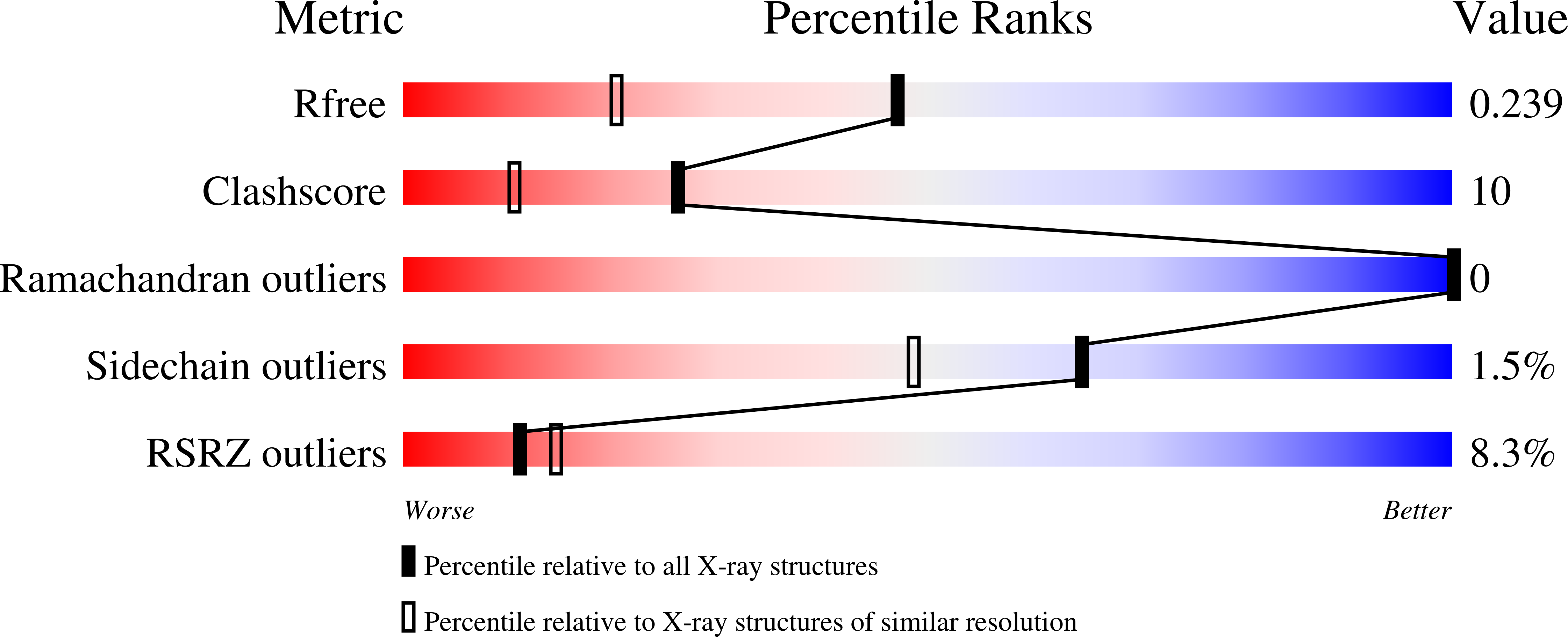
Resolution:
1.75 Å
R-Value Free:
0.24
R-Value Work:
0.20
R-Value Observed:
0.20
Space Group:
P 21 21 21
Macromolecular Entities
Polymer Type:polypeptide(L)
Molecule:adenosine kinase
Gene (Uniprot):AK
Mutations:G270S, S360F, L361T, P362S, C363G
Chain IDs:A
Chain Length:383
Number of Molecules:1
Biological Source:Toxoplasma gondii
Primary Citation
Substrate analogs induce an intermediate conformational change in Toxoplasma gondii adenosine kinase
Acta Crystallogr.,Sect.D 63 126 134 (2007)
PMID: 17242506 DOI: 10.1107/S0907444906043654

Abstact

Adenosine kinase (AK) is a key enzyme in purine metabolism in the ubiquitous intracellular parasite Toxoplasma gondii and is a potential chemotherapeutic target for the treatment of T. gondii infections. To better understand the structure-activity relationship of 6-substituted purine ribosides, the structures of the T. gondii AK-N6,N6-dimethyladenosine (DMA) complex, the AK-DMA-AMP-PCP complex, the AK-6-methyl mercaptopurine riboside (MMPR) complex and the AK-MMPR-AMP-PCP complex were determined to 1.35, 1.35, 1.75 and 1.75 A resolution, respectively. These structures reveal a conformation intermediate between open and closed, with a small lid-domain rotation of 12 degrees . Residues Gly143-X-X-Gly146 undergo torsional changes upon substrate binding, which together with a Gly68-Gly69 switch induces a hinge bending of the lid domain. The intermediate conformation suggests that ATP binding is independent of adenosine binding. Orienting the gamma-phosphate group of ATP into the optimal catalytic position may be the last step before the onset of chemical catalysis and may require the translocation of Arg136 following the complete closure of the lid domain. 6-Substituted purine-nucleoside analogs are accommodated in a hydrophobic cavity. Modification at the N6 or C6 position of the nucleoside would affect the interactions with the surrounding residues and the binding affinity.

Legend

Protein

Chemical

Disease

Primary Citation of related structures