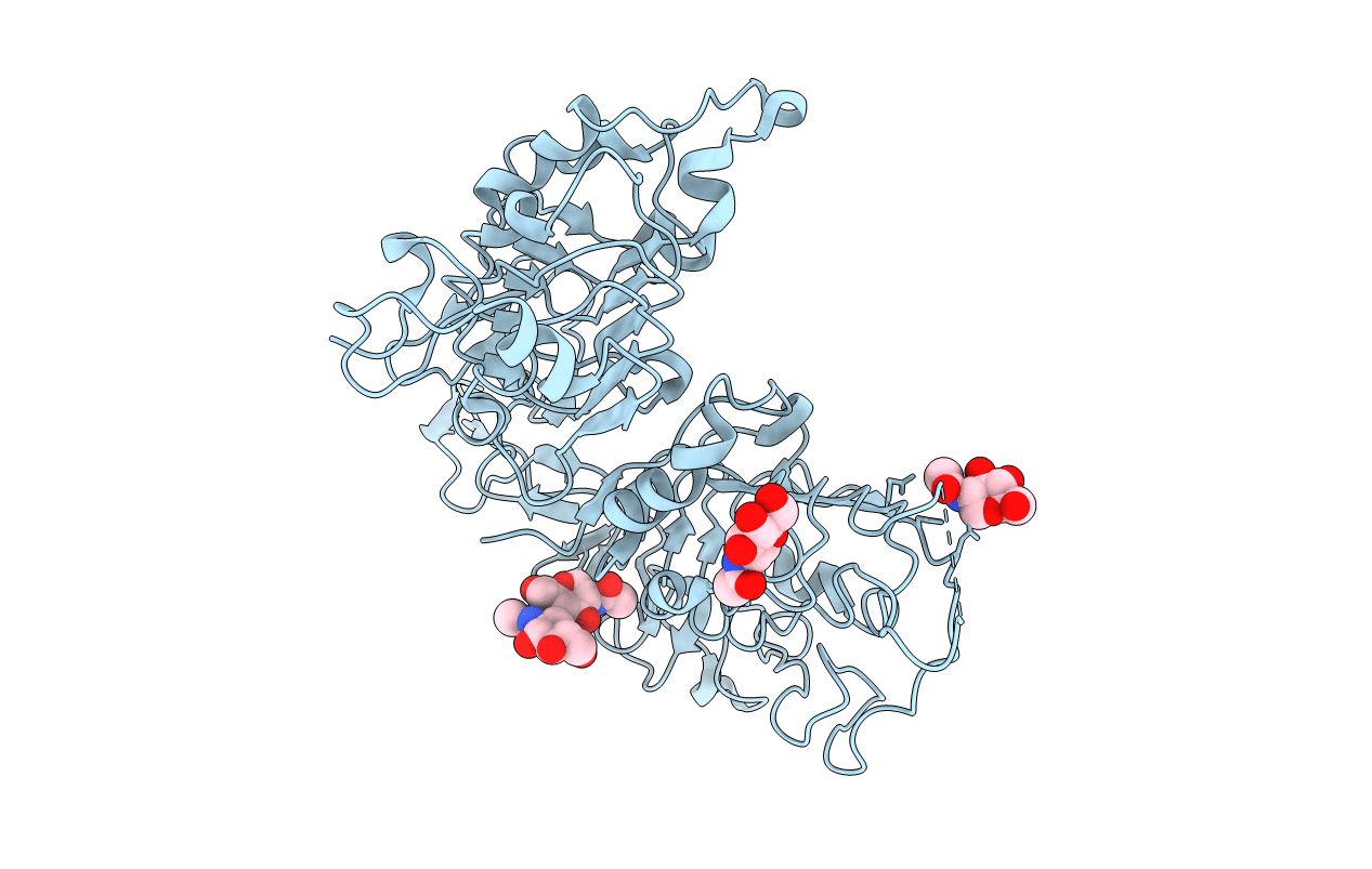
2A91 image
Entry Detail
PDB ID:
2A91
Title:
Crystal structure of ErbB2 domains 1-3
Biological Source:
Source Organism:
Host Organism:
PDB Version:
Deposition Date:
2005-07-11
Release Date:
2005-07-26
Method Details:
Experimental Method:
Resolution:
2.50 Å
R-Value Free:
0.26
R-Value Work:
0.22
R-Value Observed:
0.22
Space Group:
P 21 21 21
Macromolecular Entities
Polymer Type:polypeptide(L)
Description:Receptor tyrosine-protein kinase erbB-2
Chain IDs:A
Chain Length:517
Number of Molecules:1
Biological Source:Homo sapiens
Modified Residue
Compound ID Chain ID Parent Comp ID Details 2D Image
ASN A ASN GLYCOSYLATION SITE
Ligand Molecules
Primary Citation
The Crystal Structure of a Truncated ErbB2 Ectodomain Reveals an Active Conformation, Poised to Interact with Other ErbB Receptors
Mol.Cell 11 495 505 (2003)
PMID: 12620236 DOI: 10.1016/S1097-2765(03)00048-0

Abstact

ErbB2 does not bind ligand, yet appears to be the major signaling partner for other ErbB receptors by forming heteromeric complexes with ErbB1, ErbB3, or ErbB4. The crystal structure of residues 1-509 of ErbB2 at 2.5 A resolution reveals an activated conformation similar to that of the EGFR when complexed with ligand and very different from that seen in the unactivated forms of ErbB3 or EGFR. The structure explains the inability of ErbB2 to bind known ligands and suggests why ErbB2 fails to form homodimers. Together, the data suggest a model in which ErbB2 is already in the activated conformation and ready to interact with other ligand-activated ErbB receptors.

Legend

Protein

Chemical

Disease

Primary Citation of related structures