
Deposition Date
2005-07-08
Release Date
2006-03-28
Last Version Date
2023-08-23
Entry Detail
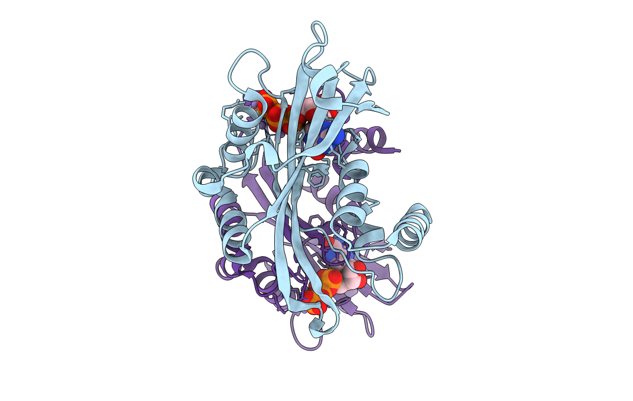
PDB ID:
2A8S
Keywords:
Title:
2.45 Angstrom Crystal Structure of the Complex Between the Nuclear SnoRNA Decapping Nudix Hydrolase X29, Manganese and GTP
Biological Source:
Source Organism(s):
Xenopus laevis (Taxon ID: 8355)
Expression System(s):
Method Details:
Experimental Method:
Resolution:
2.45 Å
R-Value Free:
0.26
R-Value Work:
0.21
Space Group:
P 21 21 21
