2A64 image
Deposition Date 2005-07-01
Release Date 2005-09-20
Last Version Date 2024-02-14
Entry Detail
PDB ID:
2A64
Keywords:
Title:
Crystal Structure of Bacterial Ribonuclease P RNA
Biological Source:
Source Organism:
(Taxon ID: )
Method Details:
Experimental Method:
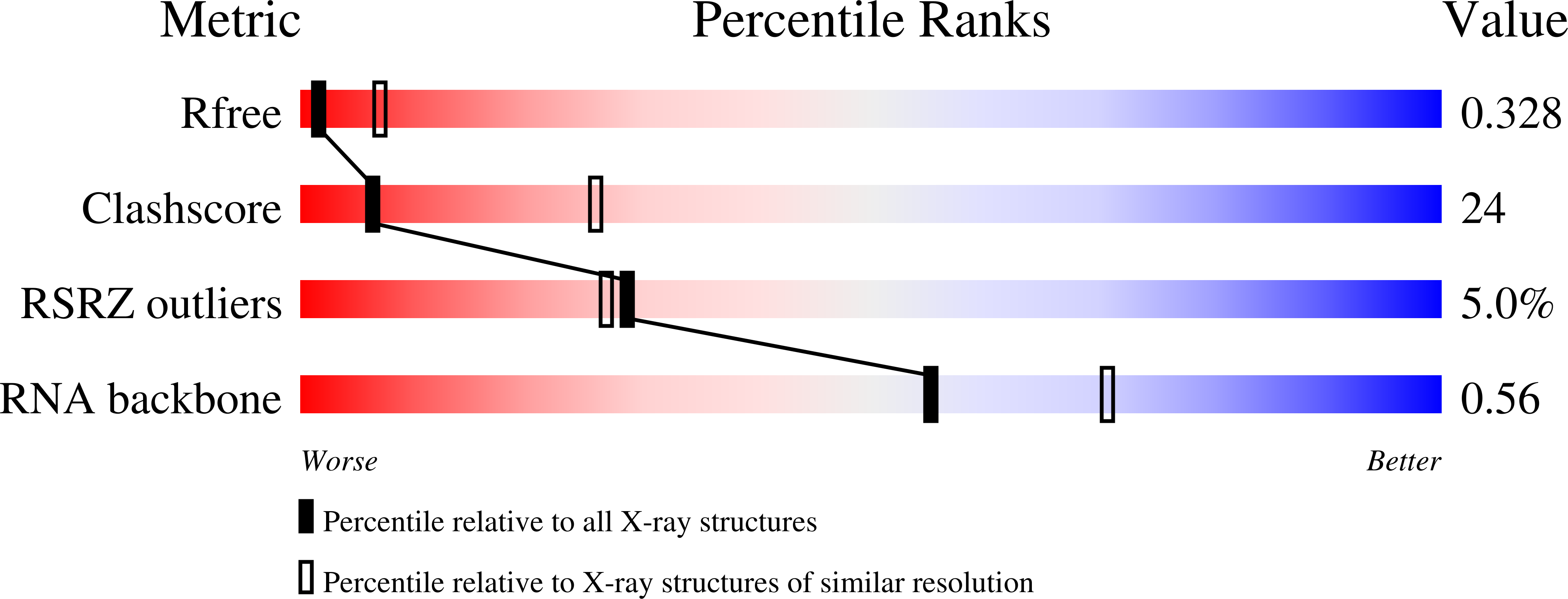
Resolution:
3.30 Å
R-Value Free:
0.33
R-Value Work:
0.32
R-Value Observed:
0.32
Space Group:
F 2 2 2
Macromolecular Entities
Polymer Type:polyribonucleotide
Molecule:ribonuclease P RNA
Mutations:G417C
Chain IDs:A
Chain Length:417
Number of Molecules:1
Biological Source:
Ligand Molecules
Primary Citation
Crystal structure of a bacterial ribonuclease P RNA.
Proc.Natl.Acad.Sci.Usa 102 13392 13397 (2005)
PMID: 16157868 DOI: 10.1073/pnas.0506662102

Abstact

The x-ray crystal structure of a 417-nt ribonuclease P RNA from Bacillus stearothermophilus was solved to 3.3-A resolution. This RNA enzyme is constructed from a number of coaxially stacked helical domains joined together by local and long-range interactions. These helical domains are arranged to form a remarkably flat surface, which is implicated by a wealth of biochemical data in the binding and cleavage of the precursors of transfer RNA substrate. Previous photoaffinity crosslinking data are used to position the substrate on the crystal structure and to identify the chemically active site of the ribozyme. This site is located in a highly conserved core structure formed by intricately interlaced long-range interactions between interhelical sequences.

Legend

Protein

Chemical

Disease

Primary Citation of related structures