2A5X image
Deposition Date 2005-07-01
Release Date 2005-08-23
Last Version Date 2023-08-23
Entry Detail
PDB ID:
2A5X
Title:
Crystal Structure of a Cross-linked Actin Dimer
Biological Source:
Source Organism:
Method Details:
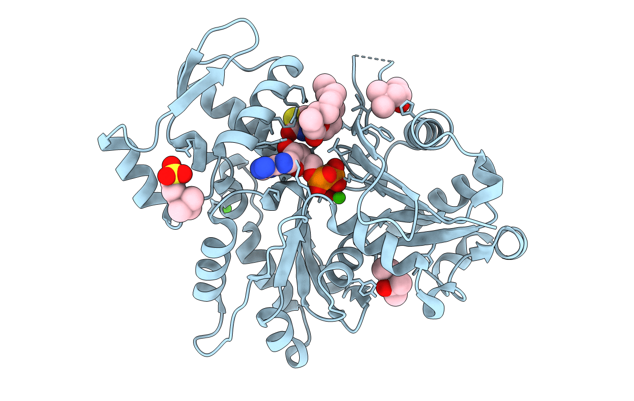
Experimental Method:
Resolution:
2.49 Å
R-Value Free:
0.25
R-Value Work:
0.19
R-Value Observed:
0.19
Space Group:
C 1 2 1
Macromolecular Entities
Structures with similar UniProt ID
Protein Blast
Polymer Type:polypeptide(L)
Molecule:Actin, alpha skeletal muscle
Gene (Uniprot):ACTA1
Chain IDs:A
Chain Length:375
Number of Molecules:1
Biological Source:Oryctolagus cuniculus
Primary Citation
The crystal structure of a cross-linked actin dimer suggests a detailed molecular interface in F-actin
Proc.Natl.Acad.Sci.Usa 102 13105 13110 (2005)
PMID: 16141336 DOI: 10.1073/pnas.0506429102

Abstact

The 2.5-A resolution crystal structure is reported for an actin dimer, composed of two protomers cross-linked along the longitudinal (or vertical) direction of the F-actin filament. The crystal structure provides an atomic resolution view of a molecular interface between actin protomers, which we argue represents a near-native interaction in the F-actin filament. The interaction involves subdomains 3 and 4 from distinct protomers. The atomic positions in the interface visualized differ by 5-10 A from those suggested by previous models of F-actin. Such differences fall within the range of uncertainties allowed by the fiber diffraction and electron microscopy methods on which previous models have been based. In the crystal, the translational arrangement of protomers lacks the slow twist found in native filaments. A plausible model of F-actin can be constructed by reintroducing the known filament twist, without disturbing significantly the interface observed in the actin dimer crystal.

Legend

Protein

Chemical

Disease

Primary Citation of related structures