2A5V image
Deposition Date 2005-07-01
Release Date 2005-09-20
Last Version Date 2023-08-23
Entry Detail
PDB ID:
2A5V
Keywords:
Title:
Crystal structure of M. tuberculosis beta carbonic anhydrase, Rv3588c, tetrameric form
Biological Source:
Source Organism:
Host Organism:
Method Details:
Experimental Method:
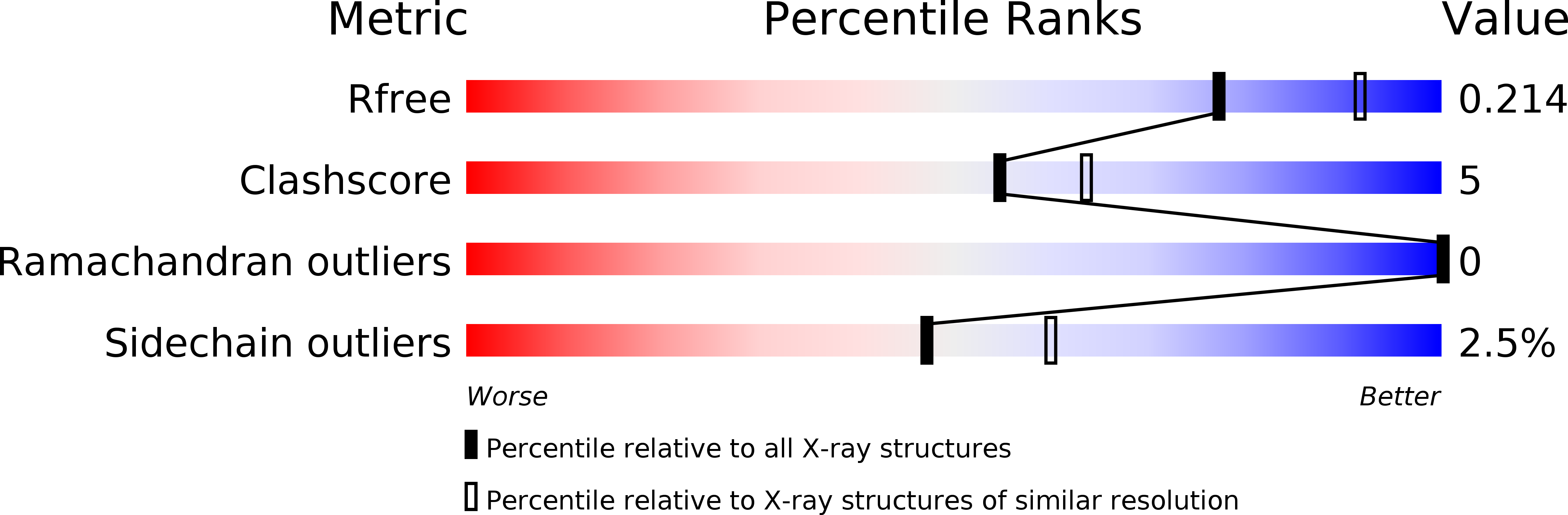
Resolution:
2.20 Å
R-Value Free:
0.21
R-Value Work:
0.16
R-Value Observed:
0.16
Space Group:
P 1 21 1
Macromolecular Entities
Structures with similar UniProt ID
Protein Blast
Polymer Type:polypeptide(L)
Molecule:CARBONIC ANHYDRASE (CARBONATE DEHYDRATASE) (CARBONIC DEHYDRATASE)
Chain IDs:A, B, C, D
Chain Length:213
Number of Molecules:4
Biological Source:Mycobacterium tuberculosis
Primary Citation
Structural Mechanics of the pH-dependent Activity of beta-Carbonic Anhydrase from Mycobacterium tuberculosis
J.Biol.Chem. 281 4993 4999 (2006)
PMID: 16321983 DOI: 10.1074/jbc.M510756200

Abstact

Carbonic anhydrases catalyze the reversible hydration of carbon dioxide to form bicarbonate, a reaction required for many functions, including carbon assimilation and pH homeostasis. Carbonic anhydrases are divided into at least three classes and are believed to share a zinc-hydroxide mechanism for carbon dioxide hydration. beta-carbonic anhydrases are broadly spread among the domains of life, and existing structures from different organisms show two distinct active site setups, one with three protein coordinations to the zinc (accessible) and the other with four (blocked). The latter is believed to be inconsistent with the zinc-hydroxide mechanism. The Mycobacterium tuberculosis Rv3588c gene, shown to be required for in vivo growth of the pathogen, encodes a beta-carbonic anhydrase with a steep pH dependence of its activity, being active at pH 8.4 but not at pH 7.5. We have recently solved the structure of this protein, which was a dimeric protein with a blocked active site. Here we present the structure of the thiocyanate complexed protein in a different crystal form. The protein now forms distinct tetramers and shows large structural changes, including a carboxylate shift yielding the accessible active site. This structure demonstrated for the first time that a beta-carbonic anhydrase can switch between the two states. A pH-dependent dimer to tetramer equilibrium was also demonstrated by dynamic light scattering measurements. The data presented here, therefore, suggest a carboxylate shift on/off switch for the enzyme, which may, in turn, be controlled by a dimer-to-tetramer equilibrium.

Legend

Protein

Chemical

Disease

Primary Citation of related structures
Feedback Form
Name
Email
Institute
Feedback