
Deposition Date
2005-06-30
Release Date
2005-07-19
Last Version Date
2024-02-14
Entry Detail
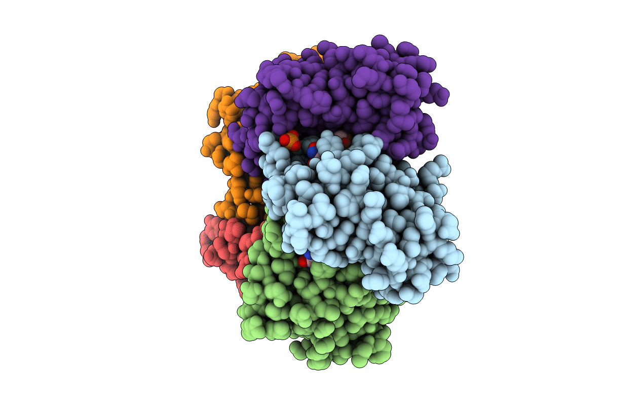
PDB ID:
2A59
Keywords:
Title:
Structure of 6,7-Dimethyl-8-ribityllumazine synthase from Schizosaccharomyces pombe mutant W27Y with bound ligand 5-nitroso-6-ribitylamino-2,4(1H,3H)-pyrimidinedione
Biological Source:
Source Organism(s):
Schizosaccharomyces pombe (Taxon ID: 4896)
Expression System(s):
Method Details:
Experimental Method:
Resolution:
2.70 Å
R-Value Free:
0.21
R-Value Work:
0.19
R-Value Observed:
0.19
Space Group:
C 2 2 21
