2A4J image
Deposition Date 2005-06-29
Release Date 2005-07-12
Last Version Date 2024-05-22
Entry Detail
PDB ID:
2A4J
Title:
Solution structure of the C-terminal domain (T94-Y172) of the human centrin 2 in complex with a 17 residues peptide (P1-XPC) from xeroderma pigmentosum group C protein
Biological Source:
Source Organism:
Homo sapiens (Taxon ID: 9606)
Method Details:
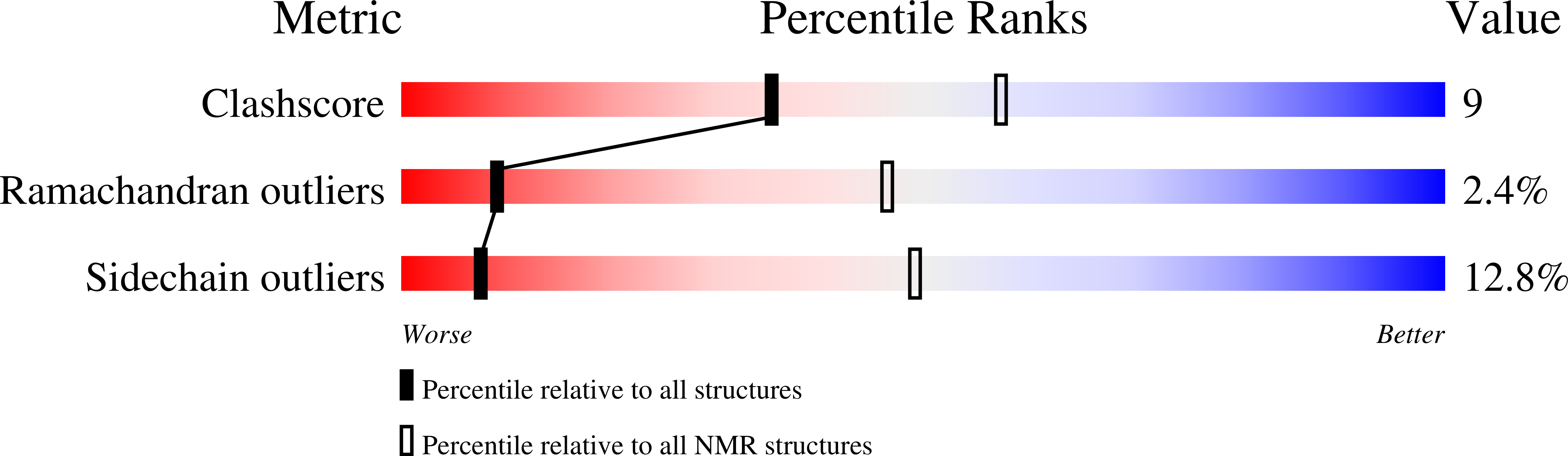
Experimental Method:
Conformers Calculated:
200
Conformers Submitted:
20
Selection Criteria:
structures with the least restraint violations
Macromolecular Entities
Structures with similar UniProt ID
Protein Blast
Polymer Type:polypeptide(L)
Molecule:Centrin 2
Gene (Uniprot):CETN2
Chain IDs:A
Chain Length:79
Number of Molecules:1
Biological Source:Homo sapiens
Structures with similar UniProt ID
Protein Blast
Polymer Type:polypeptide(L)
Molecule:17-mer peptide P1-XPC from DNA-repair protein complementing XP-C cells
Chain IDs:B
Chain Length:17
Number of Molecules:1
Biological Source:
Ligand Molecules
Primary Citation
Flexibility and plasticity of human centrin 2 binding to the xeroderma pigmentosum group C protein (XPC) from nuclear excision repair.
Biochemistry 45 3653 3663 (2006)
PMID: 16533048 DOI: 10.1021/bi0524868

Abstact

Human centrin 2 is a component of the nucleotide excision repair system, as a subunit of the heterotrimer including xeroderma pigmentosum group C protein (XPC) and hHR23B. The C-terminal domain of centrin (C-HsCen2) binds strongly a peptide from the XPC protein (P1-XPC: N(847)-R(863)). Here, we characterize the solution Ca(2+)-dependent structural and molecular features of the C-HsCen2 in complex with P1-XPC, mainly using NMR spectroscopy and molecular modeling. The N-terminal half of the peptide, organized as an alpha helix is anchored into a deep hydrophobic cavity of the protein, because of three bulky hydrophobic residues in position 1-4-8 and electrostatic contacts with the centrin helix E. Investigation of the whole centrin interactions shows that the N-terminal domain of the protein is not involved in the complex formation and is structurally independent from the peptide-bound C-terminal domain. The complex may exist in three different binding conformations corresponding to zero, one, and two Ca(2+)-bound states, which may exchange with various rates and have distinct structural stability. The various features of the intermolecular interaction presented here constitute a centrin-specific mode for the target binding.

Legend

Protein

Chemical

Disease

Primary Citation of related structures