2A45 image
Deposition Date 2005-06-27
Release Date 2006-05-02
Last Version Date 2024-11-13
Entry Detail
PDB ID:
2A45
Title:
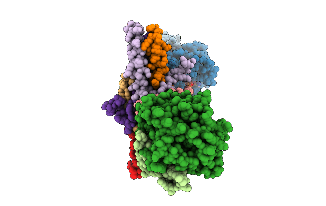
Crystal structure of the complex between thrombin and the central "E" region of fibrin
Biological Source:
Source Organism:
Homo sapiens (Taxon ID: 9606)
Method Details:
Experimental Method:
Resolution:
3.65 Å
R-Value Free:
0.29
R-Value Observed:
0.22
Space Group:
P 31
Macromolecular Entities
Polymer Type:polypeptide(L)
Molecule:Thrombin light chain
Gene (Uniprot):F2
Chain IDs:A, C (auth: D)
Chain Length:36
Number of Molecules:2
Biological Source:Homo sapiens
Polymer Type:polypeptide(L)
Molecule:Thrombin heavy chain
Gene (Uniprot):F2
Chain IDs:B, D (auth: E)
Chain Length:259
Number of Molecules:2
Biological Source:Homo sapiens
Polymer Type:polypeptide(L)
Molecule:Fibrinogen alpha chain
Gene (Uniprot):FGA
Chain IDs:E (auth: G), H (auth: J)
Chain Length:57
Number of Molecules:2
Biological Source:Homo sapiens
Polymer Type:polypeptide(L)
Molecule:Fibrinogen beta chain
Gene (Uniprot):FGB
Chain IDs:F (auth: H), I (auth: K)
Chain Length:91
Number of Molecules:2
Biological Source:Homo sapiens
Polymer Type:polypeptide(L)
Molecule:Fibrinogen gamma chain
Gene (Uniprot):FGG
Chain IDs:G (auth: I), J (auth: L)
Chain Length:45
Number of Molecules:2
Biological Source:Homo sapiens
Peptide-like Molecules
PRD_000020
Primary Citation
Structural basis for sequential cleavage of fibrinopeptides upon fibrin assembly.
Biochemistry 45 3588 3597 (2006)
PMID: 16533041 DOI: 10.1021/bi0525369

Abstact

Nonsubstrate interaction of thrombin with fibrinogen promotes sequential cleavage of fibrinopeptides A and B (fpA and fpB, respectively) from the latter, resulting in its conversion into fibrin. The recently established crystal structure of human thrombin in complex with the central part of human fibrin clarified the mechanism of this interaction. Here, we reveal new details of the structure and present the results of molecular modeling of the fpA- and fpB-containing portions of the Aalpha and Bbeta chains, not identified in the complex, in both fibrinogen and protofibrils. The analysis of the results reveals that in fibrinogen the fpA-containing portions are in a more favorable position to bind in the active site cleft of bound thrombin. Surface plasmon resonance experiments establish that the fpB-containing portions interact with the fibrin-derived dimeric D-D fragment, suggesting that in protofibrils they bind to the newly formed DD regions bringing fpB into the vicinity of bound thrombin. These findings provide a coherent rationale for the preferential removal of fpA from fibrinogen at the first stage of fibrin assembly and the accelerated cleavage of fpB from protofibrils and/or fibrils at the second stage.

Legend

Protein

Chemical

Disease

Primary Citation of related structures