2A2Z image
Entry Detail
PDB ID:
2A2Z
Keywords:
Title:
Crystal Structure of human deoxycytidine kinase in complex with deoxycytidine and uridine diphosphate
Biological Source:
Source Organism:
Host Organism:
PDB Version:
Deposition Date:
2005-06-23
Release Date:
2006-01-17
Method Details:
Experimental Method:
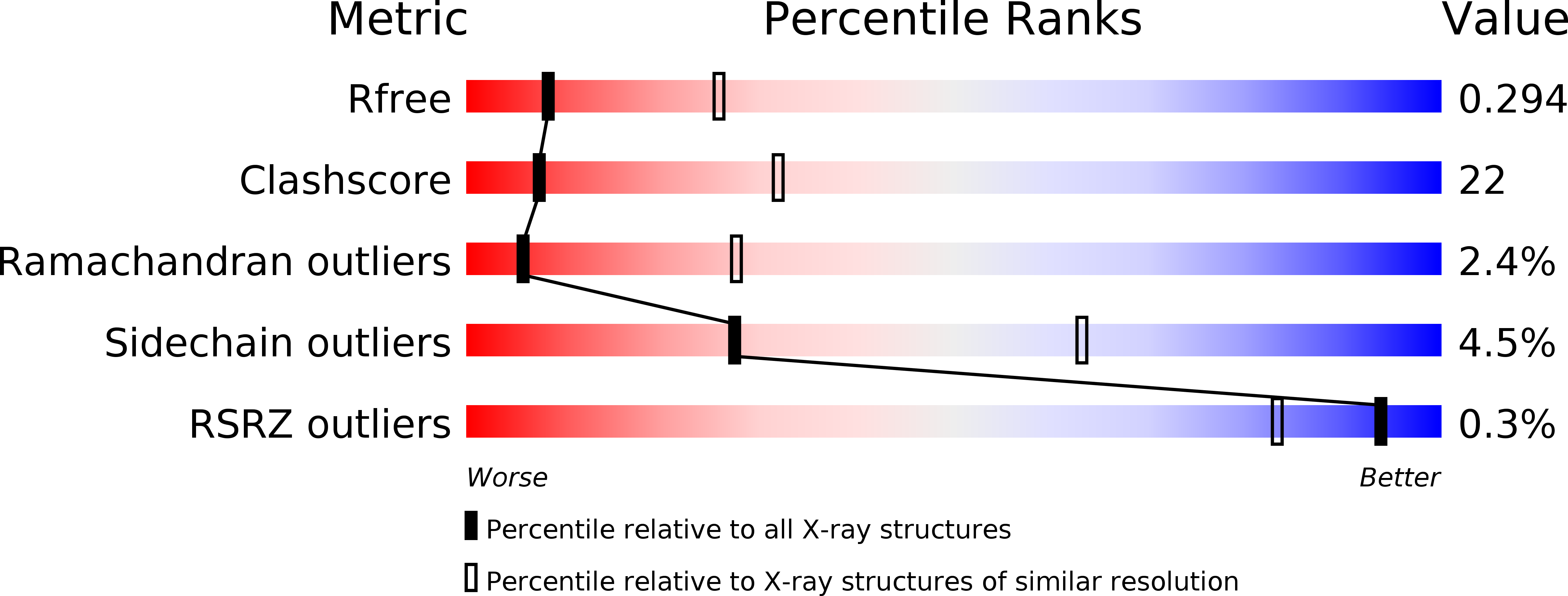
Resolution:
3.02 Å
R-Value Free:
0.3
R-Value Work:
0.24
R-Value Observed:
0.25
Space Group:
P 21 21 21
Macromolecular Entities
Polymer Type:polypeptide(L)
Description:Deoxycytidine kinase
Mutations:residues 65-79 deletion
Chain IDs:A, B, C, D
Chain Length:248
Number of Molecules:4
Biological Source:Homo sapiens
Primary Citation
Structural basis for the preference of UTP over ATP in human deoxycytidine kinase: illuminating the role of main-chain reorganization.
Biochemistry 45 452 461 (2006)
PMID: 16401075 DOI: 10.1021/bi0518646

Abstact

Human deoxycytidine kinase (dCK) uses nucleoside triphosphates to phosphorylate several clinically important prodrugs in addition to its natural substrates. Although UTP is the preferred phosphoryl donor for this reaction, our previous studies reported dCK structures solely containing ADP in the phosphoryl donor binding site. To determine the molecular basis of the kinetically observed phosphoryl donor preference, we solved crystal structures of a dCK variant lacking a flexible insert (residues 65-79) but having similar catalytic properties as wild type, in complex with deoxycytidine (dC) and UDP, and in the presence of dC but the absence of UDP or ADP. These structures reveal major changes in the donor base binding loop (residues 240-247) between the UDP-bound and ADP-bound forms, involving significant main-chain rearrangement. This loop is disordered in the dCK-dC structure, which lacks a ligand at the phosphoryl donor site. In comparison with the ADP-bound form, in the presence of UDP this loop is shifted inward to make closer contact to the smaller uracil base. These structures illuminate the phosphoryl donor binding and preference mechanisms of dCK.

Legend

Protein

Chemical

Disease

Primary Citation of related structures