
Deposition Date
2005-06-22
Release Date
2005-07-05
Last Version Date
2023-08-23
Entry Detail
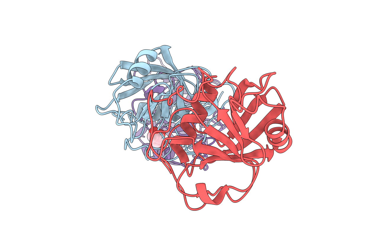
PDB ID:
2A2N
Keywords:
Title:
Crystal Structure of the peptidylprolyl isomerase domain of Human PPWD1
Biological Source:
Source Organism(s):
Homo sapiens (Taxon ID: 9606)
Expression System(s):
Method Details:
Experimental Method:
Resolution:
1.65 Å
R-Value Free:
0.24
R-Value Work:
0.19
R-Value Observed:
0.20
Space Group:
C 1 2 1
