2A2J image
Deposition Date 2005-06-22
Release Date 2005-08-23
Last Version Date 2024-10-09
Entry Detail
PDB ID:
2A2J
Keywords:
Title:
Crystal structure of a putative pyridoxine 5'-phosphate oxidase (Rv2607) from Mycobacterium tuberculosis
Biological Source:
Source Organism:
Method Details:
Experimental Method:
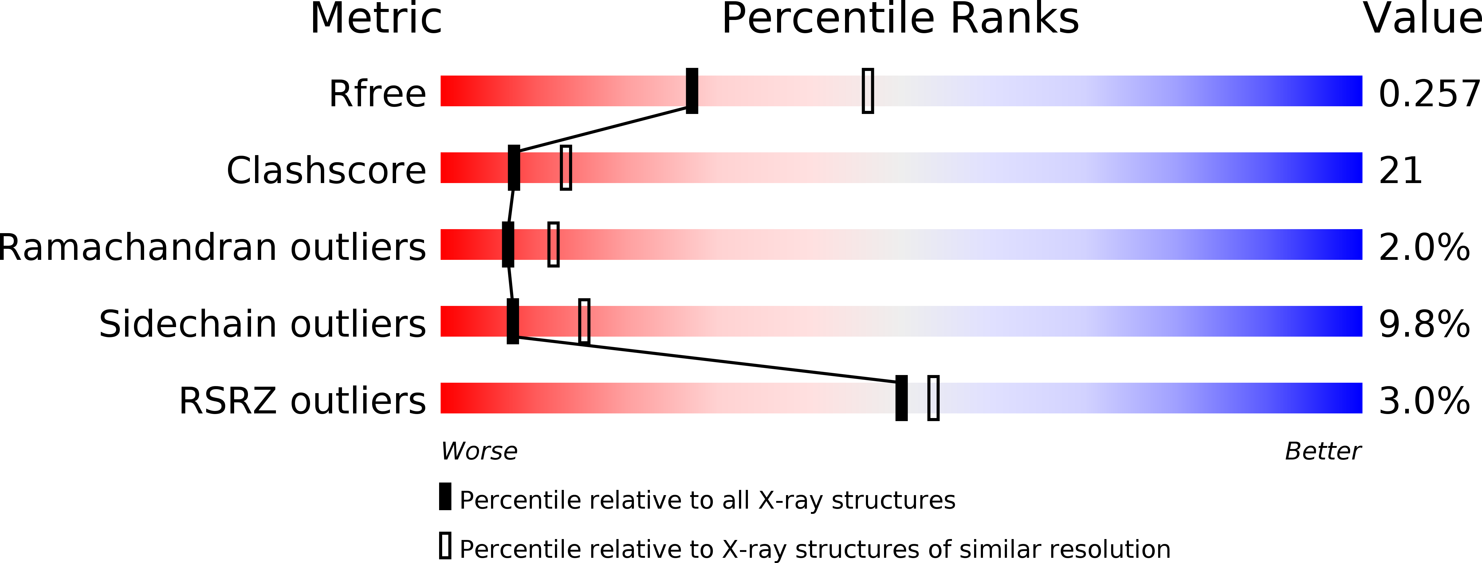
Resolution:
2.50 Å
R-Value Free:
0.26
R-Value Work:
0.20
R-Value Observed:
0.21
Space Group:
P 21 21 21
Macromolecular Entities
Structures with similar UniProt ID
Protein Blast
Polymer Type:polypeptide(L)
Molecule:Pyridoxamine 5'-phosphate oxidase
Chain IDs:A, B
Chain Length:246
Number of Molecules:2
Biological Source:Mycobacterium tuberculosis
Modified Residue
Compound ID Chain ID Parent Comp ID Details 2D Image
CME A CYS S,S-(2-HYDROXYETHYL)THIOCYSTEINE
Primary Citation
Crystal structure of a putative pyridoxine 5'-phosphate oxidase (Rv2607) from Mycobacterium tuberculosis.
Proteins 62 563 569 (2005)
PMID: 16374842 DOI: 10.1002/prot.20824

Abstact

The three-dimensional structure of Rv2607, a putative pyridoxine 5'-phosphate oxidase (PNPOx) from Mycobacterium tuberculosis, has been determined by X-ray crystallography to 2.5 A resolution. Rv2607 has a core domain similar to known PNPOx structures with a flavin mononucleotide (FMN) cofactor. Electron density for two FMN at the dimer interface is weak despite the bright yellow color of the protein solution and crystal. The shape and size of the putative binding pocket is markedly different from that of members of the PNPOx family, which may indicate some significant changes in the FMN binding mode of this protein relative to members of the family.

Legend

Protein

Chemical

Disease

Primary Citation of related structures
Feedback Form
Name
Email
Institute
Feedback