2A2C image
Deposition Date 2005-06-22
Release Date 2005-07-26
Last Version Date 2024-02-14
Entry Detail
PDB ID:
2A2C
Keywords:
Title:
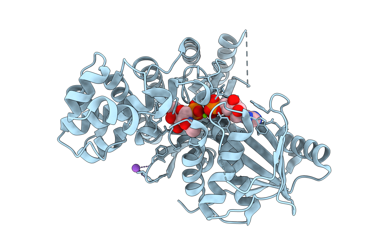
x-ray structure of human N-acetyl galactosamine kinase complexed with Mg-ADP and N-acetyl galactosamine 1-phosphate
Biological Source:
Source Organism:
Homo sapiens (Taxon ID: 9606)
Host Organism:
Method Details:
Experimental Method:
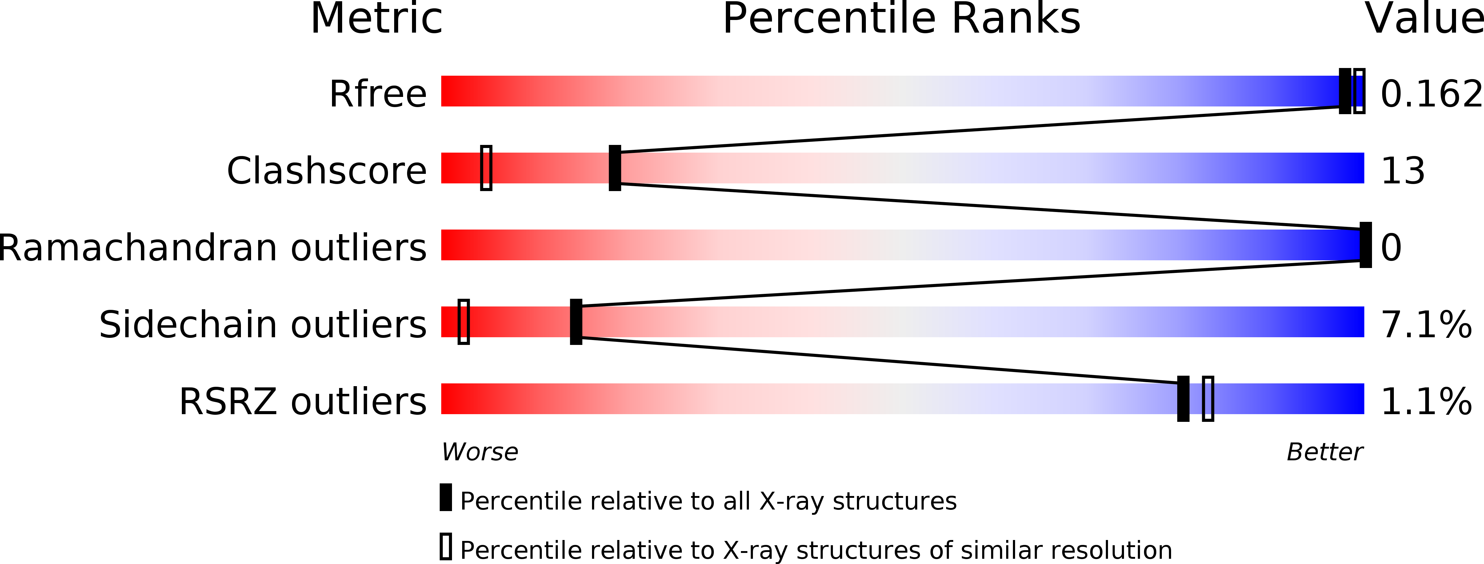
Resolution:
1.65 Å
R-Value Free:
0.20
R-Value Work:
0.16
R-Value Observed:
0.16
Space Group:
P 65
Macromolecular Entities
Structures with similar UniProt ID
Protein Blast
Polymer Type:polypeptide(L)
Molecule:N-acetylgalactosamine kinase
Gene (Uniprot):GALK2
Chain IDs:A
Chain Length:478
Number of Molecules:1
Biological Source:Homo sapiens
Primary Citation
The molecular architecture of human N-acetylgalactosamine kinase.
J.Biol.Chem. 280 32784 32791 (2005)
PMID: 16006554 DOI: 10.1074/jbc.M505730200

Abstact

Galactokinase plays a key role in normal galactose metabolism by catalyzing the conversion of alpha-d-galactose to galactose 1-phosphate. Within recent years, the three-dimensional structures of human galactokinase and two bacterial forms of the enzyme have been determined. Originally, the gene encoding galactokinase in humans was mapped to chromosome 17. An additional gene, encoding a protein with sequence similarity to galactokinase, was subsequently mapped to chromosome 15. Recent reports have shown that this second gene (GALK2) encodes an enzyme with greater activity against GalNAc than galactose. This enzyme, GalNAc kinase, has been implicated in a salvage pathway for the reutilization of free GalNAc derived from the degradation of complex carbohydrates. Here we report the first structural analysis of a GalNAc kinase. The structure of the human enzyme was solved in the presence of MnAMPPNP and GalNAc or MgATP and GalNAc (which resulted in bound products in the active site). The enzyme displays a distinctly bilobal appearance with its active site wedged between the two domains. The N-terminal region is dominated by a seven-stranded mixed beta-sheet, whereas the C-terminal motif contains two layers of anti-parallel beta-sheet. The overall topology displayed by GalNAc kinase places it into the GHMP superfamily of enzymes, which generally function as small molecule kinases. From this investigation, the geometry of the GalNAc kinase active site before and after catalysis has been revealed, and the determinants of substrate specificity have been defined on a molecular level.

Legend

Protein

Chemical

Disease

Primary Citation of related structures