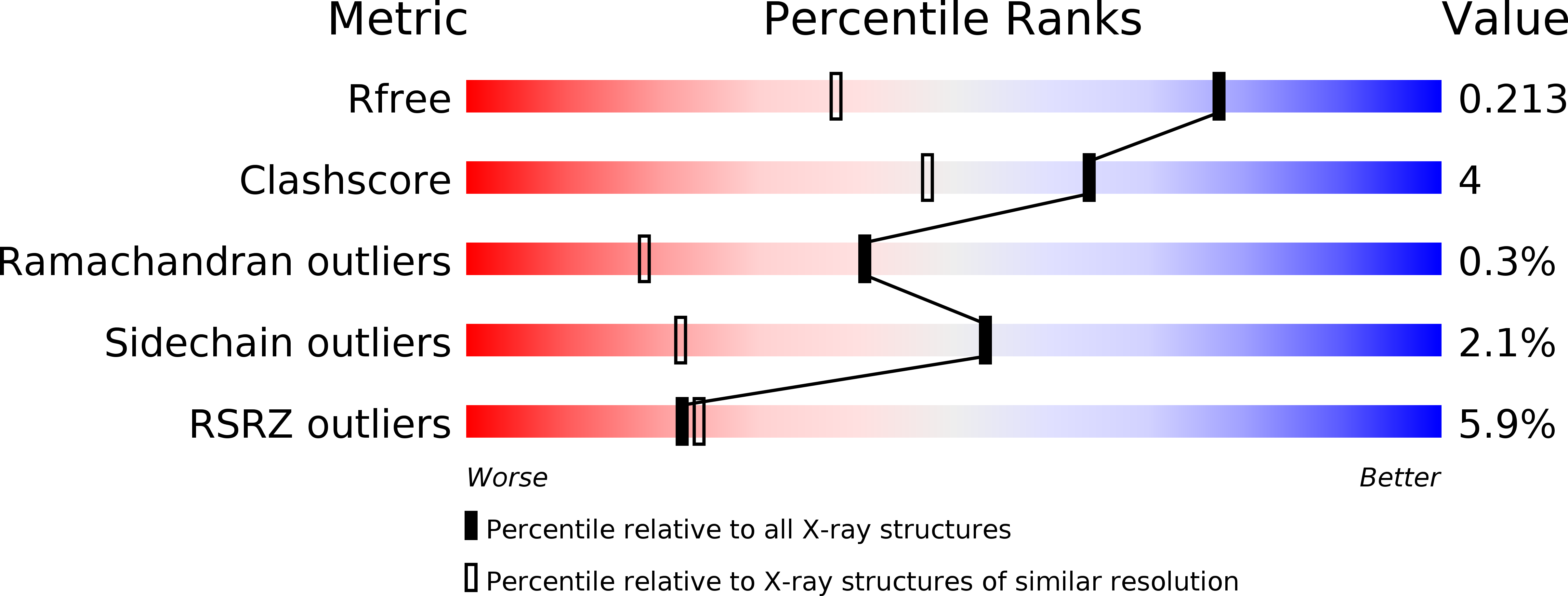
2A2A image
Deposition Date 2005-06-22
Release Date 2006-10-17
Last Version Date 2023-10-25
Entry Detail
PDB ID:
2A2A
Keywords:
Title:
High-resolution crystallographic analysis of the autoinhibited conformation of a human death-associated protein kinase
Biological Source:
Source Organism:
Homo sapiens (Taxon ID: 9606)
Host Organism:
Method Details:
Experimental Method:
Resolution:
1.47 Å
R-Value Free:
0.20
R-Value Work:
0.14
R-Value Observed:
0.15
Space Group:
P 1
Macromolecular Entities
Structures with similar UniProt ID
Protein Blast
Polymer Type:polypeptide(L)
Molecule:Death-associated protein kinase 2
Gene (Uniprot):DAPK2
Chain IDs:A, B, C, D
Chain Length:321
Number of Molecules:4
Biological Source:Homo sapiens
Primary Citation
Death-Associated Protein Kinase Activity Is Regulated by Coupled Calcium/Calmodulin Binding to Two Distinct Sites
Structure ? ? ? (2016)
PMID: 27133022 DOI: 10.1016/j.str.2016.03.020

Abstact

The regulation of many protein kinases by binding to calcium/calmodulin connects two principal mechanisms in signaling processes: protein phosphorylation and responses to dose- and time-dependent calcium signals. We used the calcium/calmodulin-dependent members of the death-associated protein kinase (DAPK) family to investigate the role of a basic DAPK signature loop near the kinase active site. In DAPK2, this loop comprises a novel dimerization-regulated calcium/calmodulin-binding site, in addition to a well-established calcium/calmodulin site in the C-terminal autoregulatory domain. Unexpectedly, impairment of the basic loop interaction site completely abolishes calcium/calmodulin binding and DAPK2 activity is reduced to a residual level, indicative of coupled binding to the two sites. This contrasts with the generally accepted view that kinase calcium/calmodulin interactions are autonomous of the kinase catalytic domain. Our data establish an intricate model of multi-step kinase activation and expand our understanding of how calcium binding connects with other mechanisms involved in kinase activity regulation.

Legend

Protein

Chemical

Disease

Primary Citation of related structures