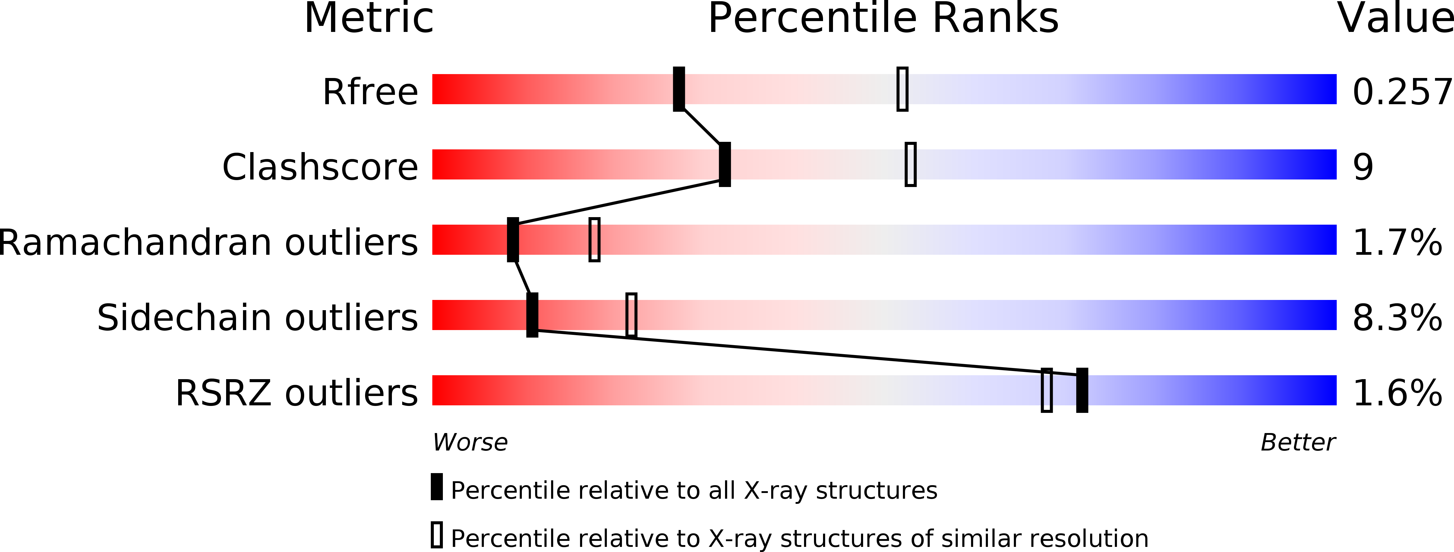
2A1S image
Deposition Date 2005-06-21
Release Date 2005-12-20
Last Version Date 2024-11-06
Entry Detail
PDB ID:
2A1S
Keywords:
Title:
Crystal structure of native PARN nuclease domain
Biological Source:
Source Organism:
Homo sapiens (Taxon ID: 9606)
Host Organism:
Method Details:
Experimental Method:
Resolution:
2.60 Å
R-Value Free:
0.25
R-Value Work:
0.21
R-Value Observed:
0.21
Space Group:
C 1 2 1
Macromolecular Entities
Polymer Type:polypeptide(L)
Molecule:Poly(A)-specific ribonuclease PARN
Gene (Uniprot):PARN
Chain IDs:A, B, C, D
Chain Length:430
Number of Molecules:4
Biological Source:Homo sapiens
Primary Citation
Structural insight into poly(A) binding and catalytic mechanism of human PARN
Embo J. 24 4082 4093 (2005)
PMID: 16281054 DOI: 10.1038/sj.emboj.7600869

Abstact

Poly(A)-specific ribonuclease (PARN) is a processive, poly(A)-specific 3' exoribonuclease. The crystal structure of C-terminal truncated human PARN determined in two states (free and RNA-bound forms) reveals that PARNn is folded into two domains, an R3H domain and a nuclease domain similar to those of Pop2p and epsilon186. The high similarity of the active site structures of PARNn and epsilon186 suggests that they may have a similar catalytic mechanism. PARNn forms a tight homodimer, with the R3H domain of one subunit partially enclosing the active site of the other subunit and poly(A) bound in a deep cavity of its nuclease domain in a sequence-nonspecific manner. The R3H domain and, possibly, the cap-binding domain are involved in poly(A) binding but these domains alone do not appear to contribute to poly(A) specificity. Mutations disrupting dimerization abolish both the enzymatic and RNA-binding activities, suggesting that the PARN dimer is a structural and functional unit. The cap-binding domain may act in concert with the R3H domain to amplify the processivity of PARN.

Legend

Protein

Chemical

Disease

Primary Citation of related structures