2A15 image
Deposition Date 2005-06-17
Release Date 2005-10-11
Last Version Date 2024-04-03
Entry Detail
PDB ID:
2A15
Title:
X-ray Crystal Structure of RV0760 from Mycobacterium Tuberculosis at 1.68 Angstrom Resolution
Biological Source:
Source Organism:
Host Organism:
Method Details:
Experimental Method:
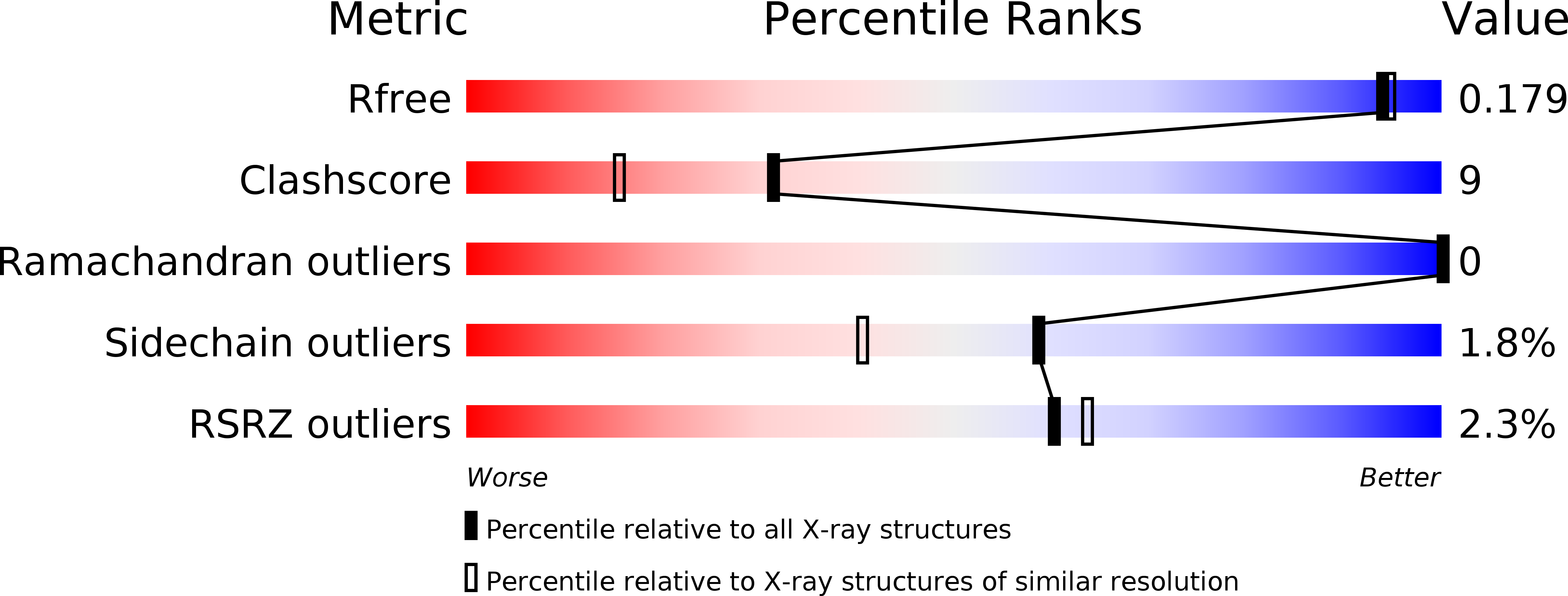
Resolution:
1.68 Å
R-Value Free:
0.20
R-Value Work:
0.17
R-Value Observed:
0.17
Space Group:
P 32 1 2
Macromolecular Entities
Polymer Type:polypeptide(L)
Molecule:HYPOTHETICAL PROTEIN Rv0760c
Gene (Uniprot):MT0785
Chain IDs:A
Chain Length:139
Number of Molecules:1
Biological Source:Mycobacterium tuberculosis
Ligand Molecules
Primary Citation
Crystal structure of Mycobacterium tuberculosis Rv0760c at 1.50 A resolution, a structural homolog of Delta(5)-3-ketosteroid isomerase
Biochim.Biophys.Acta 1784 1625 1632 (2008)
PMID: 18589008 DOI: 10.1016/j.bbapap.2008.05.012

Abstact

We have determined the X-ray crystal structure of the Mycobacterium tuberculosis (Mtb) gene product encoded by the open reading frame Rv0760c at 1.50 A resolution by single-wavelength anomalous dispersion (SAD) phasing of diffraction data from crystals of the selenomethionine-substituted protein. Refinement against diffraction data from the native protein resulted in R(work)=19.5% and R(free)=21.4%. The X-ray crystal structure shows that the homodimeric Rv0760c polypeptide has an alpha + beta conical barrel fold placing it among many structural neighbors of the nuclear transport factor 2 family (NTF2). This family is highly conserved in terms of structure; however the substrates and individual protein functions are diverse. The structures of native Rv0760c in several different crystal forms and Rv0760c bound to 17beta-estradiol 17-hemisuccinate (EH) have also been solved and analyzed.

Legend

Protein

Chemical

Disease

Primary Citation of related structures